 Movie
Movie Controller

[English] 日本語
 Yorodumi
- EMDB-1940: Negative stain EM density of green-type rubisco activase (R294V) ... -
+ Open data

- Basic information
| Entry | Database: EMDB / ID: EMD-1940 | |||||||||
|---|---|---|---|---|---|---|---|---|---|---|
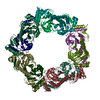
| Title | Negative stain EM density of green-type rubisco activase (R294V) from tobacco | |||||||||
 Map data Map data | Negative stain EM density of green-type rubisco activase from tobacco. Hexameric model based on p97 D2 ring is fitted into the structure. | |||||||||
 Sample Sample |
| |||||||||
 Keywords Keywords | green type rubisco activase / AAA+ protein / ATPase / negative stain EM | |||||||||
| Function / homology |  Function and homology information Function and homology informationribulose-1,5-bisphosphate carboxylase/oxygenase activator activity / thylakoid / chloroplast stroma / ATP hydrolysis activity / ATP binding / identical protein binding Similarity search - Function | |||||||||
| Biological species |  | |||||||||
| Method | single particle reconstruction / negative staining / Resolution: 20.0 Å | |||||||||
 Authors Authors | Stotz M / Mueller-Cajar O / Ciniawsky S / Wendler P / Hartl FU / Bracher A / Hayer-Hartl M | |||||||||
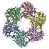
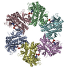
 Citation Citation | Journal: Nat Struct Mol Biol / Year: 2011 Title: Structure of green-type Rubisco activase from tobacco. Authors: Mathias Stotz / Oliver Mueller-Cajar / Susanne Ciniawsky / Petra Wendler / F Ulrich Hartl / Andreas Bracher / Manajit Hayer-Hartl /  Abstract: Rubisco, the enzyme that catalyzes the fixation of atmospheric CO(2) in photosynthesis, is subject to inactivation by inhibitory sugar phosphates. Here we report the 2.95-Å crystal structure of ...Rubisco, the enzyme that catalyzes the fixation of atmospheric CO(2) in photosynthesis, is subject to inactivation by inhibitory sugar phosphates. Here we report the 2.95-Å crystal structure of Nicotiana tabacum Rubisco activase (Rca), the enzyme that facilitates the removal of these inhibitors. Rca from tobacco has a classical AAA(+)-protein domain architecture. Although Rca populates a range of oligomeric states when in solution, it forms a helical arrangement with six subunits per turn when in the crystal. However, negative-stain electron microscopy of the active mutant R294V suggests that Rca functions as a hexamer. The residues determining species specificity for Rubisco are located in a helical insertion of the C-terminal domain and probably function in conjunction with the N-domain in Rubisco recognition. Loop segments exposed toward the central pore of the hexamer are required for the ATP-dependent remodeling of Rubisco, resulting in the release of inhibitory sugar. | |||||||||
| History |
|
- Structure visualization
| Movie |
Movie viewer |
|---|---|
| Structure viewer | EM map: SurfViewMolmilJmol/JSmol |
| Supplemental images |

- Downloads & links
-EMDB archive
| Map data | emd_1940.map.gz | 147.1 KB | EMDB map data format | |
|---|---|---|---|---|
| Header (meta data) | emd-1940-v30.xmlemd-1940.xml | 10.2 KB 10.2 KB | Display Display | EMDB header |
| Images | 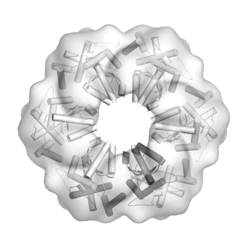 emd-1940.png emd-1940.png | 122 KB | ||
| Archive directory |  http://ftp.pdbj.org/pub/emdb/structures/EMD-1940ftp://ftp.pdbj.org/pub/emdb/structures/EMD-1940 http://ftp.pdbj.org/pub/emdb/structures/EMD-1940ftp://ftp.pdbj.org/pub/emdb/structures/EMD-1940 | HTTPS FTP |
-Related structure data
| Related structure data |  3zw6MC 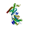 3t15C M: atomic model generated by this map C: citing same article ( |
|---|---|
| Similar structure data |


-Links
| EMDB pages | EMDB (EBI/PDBe) / EMDataResource |
|---|
-Map
| File | Download / File: emd_1940.map.gz / Format: CCP4 / Size: 7.8 MB / Type: IMAGE STORED AS FLOATING POINT NUMBER (4 BYTES) | ||||||||||||||||||||||||||||||||||||||||||||||||||||||||||||||||||||
|---|---|---|---|---|---|---|---|---|---|---|---|---|---|---|---|---|---|---|---|---|---|---|---|---|---|---|---|---|---|---|---|---|---|---|---|---|---|---|---|---|---|---|---|---|---|---|---|---|---|---|---|---|---|---|---|---|---|---|---|---|---|---|---|---|---|---|---|---|---|
| Annotation | Negative stain EM density of green-type rubisco activase from tobacco. Hexameric model based on p97 D2 ring is fitted into the structure. | ||||||||||||||||||||||||||||||||||||||||||||||||||||||||||||||||||||
| Projections & slices | Image control
Images are generated by Spider. | ||||||||||||||||||||||||||||||||||||||||||||||||||||||||||||||||||||
| Voxel size | X=Y=Z: 3.308 Å | ||||||||||||||||||||||||||||||||||||||||||||||||||||||||||||||||||||
| Density |
| ||||||||||||||||||||||||||||||||||||||||||||||||||||||||||||||||||||
| Symmetry | Space group: 1 | ||||||||||||||||||||||||||||||||||||||||||||||||||||||||||||||||||||
| Details | EMDB XML:
CCP4 map header:
| ||||||||||||||||||||||||||||||||||||||||||||||||||||||||||||||||||||
 Z (Sec.)
 Y (Row.)
 X (Col.)


-Supplemental data
- Sample components
-Entire : Nicotiana tabacum Rubisco Activase (R294V)
| Entire | Name: Nicotiana tabacum Rubisco Activase (R294V) |
|---|---|
| Components |
|
-Supramolecule #1000: Nicotiana tabacum Rubisco Activase (R294V)
| Supramolecule | Name: Nicotiana tabacum Rubisco Activase (R294V) / type: sample / ID: 1000 / Oligomeric state: Hexamer / Number unique components: 1 |
|---|---|
| Molecular weight | Experimental: 300 KDa / Theoretical: 300 KDa / Method: DSS cross linking, gel filtration |
-Macromolecule #1: Ribulose bisphosphate carboxylase activase 1, chloroplastic
| Macromolecule | Name: Ribulose bisphosphate carboxylase activase 1, chloroplastic type: protein_or_peptide / ID: 1 / Name.synonym: Rubisco Activase (Rca) Details: Hexamer assembles upon addition of 1 mM ATPgammaS at 25 deg C Number of copies: 6 / Oligomeric state: Hexamer / Recombinant expression: Yes |
|---|---|
| Source (natural) | Organism: |
| Molecular weight | Experimental: 300 KDa / Theoretical: 300 KDa |
| Recombinant expression | Organism:  |
| Sequence | InterPro: ATPase, AAA-type, core |
-Experimental details
-Structure determination
| Method | negative staining |
|---|---|
 Processing Processing | single particle reconstruction |
| Aggregation state | particle |
-Sample preparation
| Concentration | 0.044 mg/mL |
|---|---|
| Buffer | pH: 8 / Details: 20mM Tris-HCl, 50mM NaCl, 2mM MgCl2,1mM ATPgammaS |
| Staining | Type: NEGATIVE Details: Grids were stained twice with 2% w/v uranyl acetate |
| Grid | Details: Plain carbon |
| Vitrification | Cryogen name: NONE / Instrument: OTHER |
- Electron microscopy
| Microscope | FEI TECNAI 12 |
|---|---|
| Alignment procedure | Legacy - Astigmatism: Objective lens astigmatism was corrected for at 110k magnification |
| Date | Jun 22, 2011 |
| Image recording | Category: CCD / Film or detector model: FEI EAGLE (2k x 2k) / Average electron dose: 20 e/Å2 |
| Electron beam | Acceleration voltage: 120 kV / Electron source: LAB6 |
| Electron optics | Illumination mode: FLOOD BEAM / Imaging mode: BRIGHT FIELD / Cs: 2.0 mm / Nominal defocus max: 0.95 µm / Nominal defocus min: 0.47 µm |
| Sample stage | Specimen holder: Single tilt / Specimen holder model: SIDE ENTRY, EUCENTRIC |
-Image processing
| CTF correction | Details: Phase flipping, each particle |
|---|---|
| Final reconstruction | Applied symmetry - Point group: C6 (6 fold cyclic) / Algorithm: OTHER / Resolution.type: BY AUTHOR / Resolution: 20.0 Å / Resolution method: FSC 0.5 CUT-OFF / Software - Name: MRC, IMAGIC, SPIDER / Number images used: 599 |
-Atomic model buiding 1
| Initial model | PDB ID: |
|---|---|
| Software | Name:  Chimera Chimera |
| Details | Protocol: Rigid body. A module of alpha helical domain and alpha-beta domain of neighbouring subunit was overlaid with p97 D2 (3CF3) structure and the hexamer was fitted into the EM map |
| Refinement | Space: REAL / Protocol: RIGID BODY FIT / Target criteria: Local minimisation, fit in map |
| Output model | PDB-3zw6: |
