[English] 日本語
Yorodumi
- PDB-3t15: Structure of green-type Rubisco activase from tobacco -

+
Open data


ID or keywords:

Loading...

-
Basic information

Entry
Database: PDB / ID: 3t15
TitleStructure of green-type Rubisco activase from tobacco
ComponentsRibulose bisphosphate carboxylase/oxygenase activase 1, chloroplastic
KeywordsPHOTOSYNTHESIS / Rubisco activase / AAA+ protein
Function / homology
Function and homology information


ribulose-1,5-bisphosphate carboxylase/oxygenase activator activity / chloroplast stroma / ATP hydrolysis activity / ATP binding / identical protein binding
Similarity search - Function
Helicase, Ruva Protein; domain 3 - #1070 / Ribulose bisphosphate carboxylase/oxygenase activase-like / : / Ribulose bisphosphate carboxylase/oxygenase activase, AAA, helical / Helicase, Ruva Protein; domain 3 / ATPase family associated with various cellular activities (AAA) / ATPase, AAA-type, core / P-loop containing nucleotide triphosphate hydrolases / Rossmann fold / P-loop containing nucleoside triphosphate hydrolase ...Helicase, Ruva Protein; domain 3 - #1070 / Ribulose bisphosphate carboxylase/oxygenase activase-like / : / Ribulose bisphosphate carboxylase/oxygenase activase, AAA, helical / Helicase, Ruva Protein; domain 3 / ATPase family associated with various cellular activities (AAA) / ATPase, AAA-type, core / P-loop containing nucleotide triphosphate hydrolases / Rossmann fold / P-loop containing nucleoside triphosphate hydrolase / Orthogonal Bundle / 3-Layer(aba) Sandwich / Mainly Alpha / Alpha Beta
Similarity search - Domain/homology
Ribulose bisphosphate carboxylase/oxygenase activase 1, chloroplastic
Similarity search - Component
Biological speciesNicotiana tabacum (common tobacco)
MethodX-RAY DIFFRACTION / SYNCHROTRON / SIRAS / Resolution: 2.95 Å
AuthorsStotz, M. / Wendler, P. / Mueller-Cajar, O. / Hartl, F.U. / Bracher, A. / Hayer-Hartl, M.
Citation
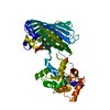
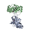
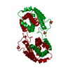
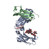
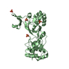
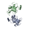
Journal: Nat Struct Mol Biol / Year: 2011
Title: Structure of green-type Rubisco activase from tobacco.
Authors: Mathias Stotz / Oliver Mueller-Cajar / Susanne Ciniawsky / Petra Wendler / F Ulrich Hartl / Andreas Bracher / Manajit Hayer-Hartl /
Abstract: Rubisco, the enzyme that catalyzes the fixation of atmospheric CO(2) in photosynthesis, is subject to inactivation by inhibitory sugar phosphates. Here we report the 2.95-Å crystal structure of ...Rubisco, the enzyme that catalyzes the fixation of atmospheric CO(2) in photosynthesis, is subject to inactivation by inhibitory sugar phosphates. Here we report the 2.95-Å crystal structure of Nicotiana tabacum Rubisco activase (Rca), the enzyme that facilitates the removal of these inhibitors. Rca from tobacco has a classical AAA(+)-protein domain architecture. Although Rca populates a range of oligomeric states when in solution, it forms a helical arrangement with six subunits per turn when in the crystal. However, negative-stain electron microscopy of the active mutant R294V suggests that Rca functions as a hexamer. The residues determining species specificity for Rubisco are located in a helical insertion of the C-terminal domain and probably function in conjunction with the N-domain in Rubisco recognition. Loop segments exposed toward the central pore of the hexamer are required for the ATP-dependent remodeling of Rubisco, resulting in the release of inhibitory sugar.
History
DepositionJul 21, 2011Deposition site: RCSB / Processing site: RCSB
Revision 1.0Nov 9, 2011Provider: repository / Type: Initial release
Revision 1.1Nov 23, 2011Group: Database references
Revision 1.2Dec 28, 2011Group: Database references
Revision 1.3Nov 20, 2019Group: Database references / Category: pdbx_database_related / Item: _pdbx_database_related.content_type
Revision 1.4Dec 18, 2019Group: Database references / Category: pdbx_database_related / Item: _pdbx_database_related.db_id
Revision 1.5Feb 28, 2024Group: Data collection / Database references / Category: chem_comp_atom / chem_comp_bond / database_2
Item: _database_2.pdbx_DOI / _database_2.pdbx_database_accession

-
Structure visualization

Structure viewerMolecule:
MolmilJmol/JSmol

Downloads & links

-
Assembly

Deposited unit
A: Ribulose bisphosphate carboxylase/oxygenase activase 1, chloroplastic


Theoretical massNumber of molelcules
Total (without water)32,8571
Polymers32,8571
Non-polymers00
Water00
1


  • Idetical with deposited unit
  • defined by software
TypeNameSymmetry operationNumber
identity operation1_555x,y,z1
MethodPISA
Unit cell
Length a, b, c (Å)103.635, 103.635, 56.675
Angle α, β, γ (deg.)90.000, 90.000, 120.000
Int Tables number170
Space group name H-MP65
DetailsTHE ANALYSIS OF THE CBBX PROTEIN IN SOLUTION AND EM STUDIES SUGGEST THAT THE BIOLOGICALLY ACTIVE OLIGOMER IS A HEXAMER, BUT IT CANNOT BE GENERATED BY THE APPLICATION OF SYMMETRY OPERATORS TO THE CHAINS IN THE COORDINATE FILE.

-
Components

#1: Protein Ribulose bisphosphate carboxylase/oxygenase activase 1, chloroplastic / RA 1 / RuBisCO activase 1


Mass: 32856.801 Da / Num. of mol.: 1
Source method: isolated from a genetically manipulated source
Source: (gene. exp.) Nicotiana tabacum (common tobacco) / Gene: Rca1 / Plasmid: pHUE / Production host: Escherichia coli (E. coli) / Strain (production host): BL21(DE3) / References: UniProt: Q40460

-
Experimental details

-
Experiment

ExperimentMethod: X-RAY DIFFRACTION / Number of used crystals: 1

-
Sample preparation

CrystalDensity Matthews: 2.67 Å3/Da / Density % sol: 54 %
Crystal growTemperature: 291 K / Method: vapor diffusion / pH: 6
Details: 50 mM MES-Na pH 6.0 and 350 mM magnesium formate, vapor diffusion, temperature 291K

-
Data collection

DiffractionMean temperature: 100 K
Diffraction sourceSource: SYNCHROTRON / Site: ESRF / Beamline: ID14-4 / Wavelength: 1.0332 Å
DetectorType: ADSC QUANTUM 315r / Detector: CCD / Date: Dec 12, 2010
RadiationProtocol: SINGLE WAVELENGTH / Monochromatic (M) / Laue (L): M / Scattering type: x-ray
Radiation wavelengthWavelength: 1.0332 Å / Relative weight: 1
ReflectionRedundancy: 5.5 % / Av σ(I) over netI: 9.7 / Number: 29140 / Rmerge(I) obs: 0.053 / Rsym value: 0.053 / D res high: 3.312 Å / D res low: 47.946 Å / Num. obs: 5298 / % possible obs: 99.7
Diffraction reflection shell
Highest resolution (Å)Lowest resolution (Å)% possible obs (%)IDRmerge(I) obsRsym valueRedundancy
10.4747.9598.510.0290.0295.1
7.4110.4710010.0270.0275.3
6.057.4110010.0370.0375.5
5.246.0510010.040.045.5
4.685.2410010.040.045.6
4.284.6810010.0530.0535.6
3.964.2810010.0970.0975.6
3.73.9610010.1840.1845.6
3.493.710010.3850.3855.7
3.313.4998.110.6120.6125.3
ReflectionResolution: 2.949→51.848 Å / Num. all: 7453 / Num. obs: 7453 / % possible obs: 99.9 % / Observed criterion σ(F): 0 / Observed criterion σ(I): 0 / Redundancy: 4 % / Rmerge(I) obs: 0.062 / Rsym value: 0.062 / Net I/σ(I): 14.6
Reflection shell

Diffraction-ID: 1

Resolution (Å)Redundancy (%)Rmerge(I) obsMean I/σ(I) obsNum. measured allNum. unique allRsym value% possible all
2.95-3.113.70.5051.5396010650.505100
3.11-3.33.70.2473.2382210220.24799.9
3.3-3.533.70.1136.635169520.113100
3.53-3.813.70.0749.733479060.074100
3.81-4.173.60.05711.329908220.057100
4.17-4.663.70.04911.627797560.049100
4.66-5.393.60.04811.424106700.048100
5.39-6.63.50.0481219915620.04899.8
6.6-9.338.30.0628.736904450.062100
9.33-56.7256.20.0618.615642530.06198.6

-
Phasing

PhasingMethod: SIRAS
Phasing MAD set site
IDCartn x (Å)Cartn y (Å)Cartn z (Å)Atom type symbolB isoOccupancy
133.31646.837.503S201
226.38251.369-1.644S200.824
346.36242.132-12.373S200.8
445.25762.2230.206S200.75
554.11861.8174.747S200.583
631.86451.4934.48S200.572
764.85158.21.362S200.501
851.49559.3927.031S200.493
928.19441.5061.624S200.492
1036.42249.869-15.204S200.384
1168.11766.6273.479S200.312
1232.88340.739.432S200.3
1359.28361.0267.951S200.26
1436.74452.20617.08S200.248
1534.84727.746-0.415S200.161
1658.46473.6933.268S200.11
1733.04751.8469.481S200.05
Phasing dmFOM : 0.61 / FOM acentric: 0.6 / FOM centric: 0.61 / Reflection: 5404 / Reflection acentric: 4954 / Reflection centric: 450
Phasing dm shell
Resolution (Å)FOM FOM acentricFOM centricReflectionReflection acentricReflection centric
9.4-47.9230.980.980.9924819454
5.9-9.40.920.930.8672964485
4.7-5.90.850.860.7690982881
4.1-4.70.730.730.6991985267
3.5-4.10.440.450.3216071499108
3.3-3.50.20.210.1299293755

-
Processing

Software
NameVersionClassificationNB
SCALA3.2.25data scaling
SHELXphasing
RESOLVE2.13phasing
REFMACrefinement
PDB_EXTRACT3.1data extraction
XDSdata reduction
SHELXDphasing
RefinementMethod to determine structure: SIRAS / Resolution: 2.95→20 Å / Cor.coef. Fo:Fc: 0.953 / Cor.coef. Fo:Fc free: 0.899 / WRfactor Rfree: 0.3269 / WRfactor Rwork: 0.2716 / Occupancy max: 1 / Occupancy min: 1 / FOM work R set: 0.761 / SU B: 47.761 / SU ML: 0.406 / SU R Cruickshank DPI: 0.3741 / SU Rfree: 0.4735 / Cross valid method: THROUGHOUT / σ(F): 0 / ESU R Free: 0.463 / Stereochemistry target values: MAXIMUM LIKELIHOOD / Details: HYDROGENS HAVE BEEN ADDED IN THE RIDING POSITIONS
RfactorNum. reflection% reflectionSelection details
Rfree0.2962 350 4.7 %RANDOM
Rwork0.226 ---
obs0.2292 7051 99.93 %-
all-7056 --
Solvent computationIon probe radii: 0.8 Å / Shrinkage radii: 0.8 Å / VDW probe radii: 1.2 Å / Solvent model: MASK
Displacement parametersBiso max: 210.76 Å2 / Biso mean: 117.6419 Å2 / Biso min: 80.07 Å2
Baniso -1Baniso -2Baniso -3
1-1.81 Å20.9 Å20 Å2
2--1.81 Å20 Å2
3----2.71 Å2
Refinement stepCycle: LAST / Resolution: 2.95→20 Å
ProteinNucleic acidLigandSolventTotal
Num. atoms1919 0 0 0 1919
Refine LS restraints
Refine-IDTypeDev idealDev ideal targetNumber
X-RAY DIFFRACTIONr_bond_refined_d0.0140.0221954
X-RAY DIFFRACTIONr_angle_refined_deg1.4491.9592657
X-RAY DIFFRACTIONr_dihedral_angle_1_deg6.815258
X-RAY DIFFRACTIONr_dihedral_angle_2_deg41.05924.74478
X-RAY DIFFRACTIONr_dihedral_angle_3_deg21.92215285
X-RAY DIFFRACTIONr_dihedral_angle_4_deg14.891159
X-RAY DIFFRACTIONr_chiral_restr0.0910.2307
X-RAY DIFFRACTIONr_gen_planes_refined0.0040.021491
X-RAY DIFFRACTIONr_nbd_refined0.2670.2985
X-RAY DIFFRACTIONr_nbtor_refined0.3220.21362
X-RAY DIFFRACTIONr_xyhbond_nbd_refined0.1430.263
X-RAY DIFFRACTIONr_symmetry_vdw_refined0.1920.236
X-RAY DIFFRACTIONr_symmetry_hbond_refined0.2280.21
X-RAY DIFFRACTIONr_mcbond_it0.4351.51330
X-RAY DIFFRACTIONr_mcangle_it0.77122058
X-RAY DIFFRACTIONr_scbond_it1.1853705
X-RAY DIFFRACTIONr_scangle_it1.9834.5599
LS refinement shellResolution: 2.95→3.025 Å / Total num. of bins used: 20
RfactorNum. reflection% reflection
Rfree0.395 27 -
Rwork0.288 494 -
all-521 -
obs--100 %
Refinement TLS params.

Method: refined / Refine-ID: X-RAY DIFFRACTION

IDL112)L122)L132)L222)L232)L332)S11 (Å °)S12 (Å °)S13 (Å °)S21 (Å °)S22 (Å °)S23 (Å °)S31 (Å °)S32 (Å °)S33 (Å °)T112)T122)T132)T222)T232)T332)Origin x (Å)Origin y (Å)Origin z (Å)
132.3803-8.23458.43072.7121.137119.6165-0.40320.43641.6888-0.33940.0345-3.2018-0.78910.47210.36870.0811-0.0606-0.1412-0.10780.13460.307930.841735.4545-4.7065
26.154-2.5421.21795.1663-0.91794.8272-0.6501-1.0044-0.46341.14170.31790.42270.5383-0.56940.33220.19370.092-0.05910.1573-0.0488-0.194113.914532.15443.3552
317.113112.027611.68279.42765.786714.0082-0.6504-0.5978-0.90080.93620.33793.68360.1448-1.38140.31250.56120.05550.36370.83350.04860.19791.224228.83788.7897
43.5576-1.52521.37843.3928-2.27923.4250.15690.33460.03530.0312-0.4423-0.66080.24950.19970.28540.11470.059-0.0557-0.07930.0866-0.081341.230214.4512-3.6981
Refinement TLS group
IDRefine-IDRefine TLS-IDAuth asym-IDAuth seq-ID
1X-RAY DIFFRACTION1A68 - 79
2X-RAY DIFFRACTION2A80 - 141
3X-RAY DIFFRACTION2A145 - 176
4X-RAY DIFFRACTION2A219 - 234
5X-RAY DIFFRACTION2A237 - 252
6X-RAY DIFFRACTION3A191 - 207
7X-RAY DIFFRACTION4A253 - 360

+
About Yorodumi

-
News

-
Feb 9, 2022. New format data for meta-information of EMDB entries

New format data for meta-information of EMDB entries

  • Version 3 of the EMDB header file is now the official format.
  • The previous official version 1.9 will be removed from the archive.

Related info.:EMDB header

External links:wwPDB to switch to version 3 of the EMDB data model

-
Aug 12, 2020. Covid-19 info

Covid-19 info

URL: https://pdbj.org/emnavi/covid19.php

New page: Covid-19 featured information page in EM Navigator.

Related info.:Covid-19 info / Mar 5, 2020. Novel coronavirus structure data

+
Mar 5, 2020. Novel coronavirus structure data

Novel coronavirus structure data

Related info.:Yorodumi Speices / Aug 12, 2020. Covid-19 info

External links:COVID-19 featured content - PDBj / Molecule of the Month (242):Coronavirus Proteases

+
Jan 31, 2019. EMDB accession codes are about to change! (news from PDBe EMDB page)

EMDB accession codes are about to change! (news from PDBe EMDB page)

  • The allocation of 4 digits for EMDB accession codes will soon come to an end. Whilst these codes will remain in use, new EMDB accession codes will include an additional digit and will expand incrementally as the available range of codes is exhausted. The current 4-digit format prefixed with “EMD-” (i.e. EMD-XXXX) will advance to a 5-digit format (i.e. EMD-XXXXX), and so on. It is currently estimated that the 4-digit codes will be depleted around Spring 2019, at which point the 5-digit format will come into force.
  • The EM Navigator/Yorodumi systems omit the EMD- prefix.

Related info.:Q: What is EMD? / ID/Accession-code notation in Yorodumi/EM Navigator

External links:EMDB Accession Codes are Changing Soon! / Contact to PDBj

+
Jul 12, 2017. Major update of PDB

Major update of PDB

  • wwPDB released updated PDB data conforming to the new PDBx/mmCIF dictionary.
  • This is a major update changing the version number from 4 to 5, and with Remediation, in which all the entries are updated.
  • In this update, many items about electron microscopy experimental information are reorganized (e.g. em_software).
  • Now, EM Navigator and Yorodumi are based on the updated data.

External links:wwPDB Remediation / Enriched Model Files Conforming to OneDep Data Standards Now Available in the PDB FTP Archive

-
Yorodumi

Thousand views of thousand structures

  • Yorodumi is a browser for structure data from EMDB, PDB, SASBDB, etc.
  • This page is also the successor to EM Navigator detail page, and also detail information page/front-end page for Omokage search.
  • The word "yorodu" (or yorozu) is an old Japanese word meaning "ten thousand". "mi" (miru) is to see.

Related info.:EMDB / PDB / SASBDB / Comparison of 3 databanks / Yorodumi Search / Aug 31, 2016. New EM Navigator & Yorodumi / Yorodumi Papers / Jmol/JSmol / Function and homology information / Changes in new EM Navigator and Yorodumi

Read more