 Movie
Movie Controller

[English] 日本語
 Yorodumi
- PDB-3zw6: MODEL OF HEXAMERIC AAA DOMAIN ARRANGEMENT OF GREEN-TYPE RUBISCO A... -
+ Open data

- Basic information
| Entry | Database: PDB / ID: 3zw6 | ||||||
|---|---|---|---|---|---|---|---|
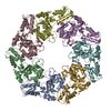
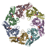
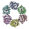
| Title | MODEL OF HEXAMERIC AAA DOMAIN ARRANGEMENT OF GREEN-TYPE RUBISCO ACTIVASE FROM TOBACCO. | ||||||
 Components Components | RIBULOSE BISPHOSPHATE CARBOXYLASE/OXYGENASE ACTIVASE 1, CHLOROPLASTIC | ||||||
 Keywords Keywords | PHOTOSYNTHESIS / NEGATIVE STAIN EM | ||||||
| Function / homology |  Function and homology information Function and homology informationribulose-1,5-bisphosphate carboxylase/oxygenase activator activity / chloroplast stroma / ATP hydrolysis activity / ATP binding / identical protein binding Similarity search - Function | ||||||
| Biological species |  | ||||||
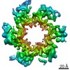
| Method | ELECTRON MICROSCOPY / single particle reconstruction / negative staining / Resolution: 20 Å | ||||||
 Authors Authors | Stotz, M. / Mueller-Cajar, O. / Ciniawsky, S. / Wendler, P. / Hartl, F.U. / Bracher, A. / Hayer-Hartl, M. | ||||||
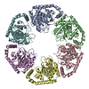
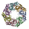
 Citation Citation | Journal: Nat Struct Mol Biol / Year: 2011 Title: Structure of green-type Rubisco activase from tobacco. Authors: Mathias Stotz / Oliver Mueller-Cajar / Susanne Ciniawsky / Petra Wendler / F Ulrich Hartl / Andreas Bracher / Manajit Hayer-Hartl /  Abstract: Rubisco, the enzyme that catalyzes the fixation of atmospheric CO(2) in photosynthesis, is subject to inactivation by inhibitory sugar phosphates. Here we report the 2.95-Å crystal structure of ...Rubisco, the enzyme that catalyzes the fixation of atmospheric CO(2) in photosynthesis, is subject to inactivation by inhibitory sugar phosphates. Here we report the 2.95-Å crystal structure of Nicotiana tabacum Rubisco activase (Rca), the enzyme that facilitates the removal of these inhibitors. Rca from tobacco has a classical AAA(+)-protein domain architecture. Although Rca populates a range of oligomeric states when in solution, it forms a helical arrangement with six subunits per turn when in the crystal. However, negative-stain electron microscopy of the active mutant R294V suggests that Rca functions as a hexamer. The residues determining species specificity for Rubisco are located in a helical insertion of the C-terminal domain and probably function in conjunction with the N-domain in Rubisco recognition. Loop segments exposed toward the central pore of the hexamer are required for the ATP-dependent remodeling of Rubisco, resulting in the release of inhibitory sugar. | ||||||
| History |
|
- Structure visualization
| Movie |
Movie viewer |
|---|---|
| Structure viewer | Molecule: MolmilJmol/JSmol |
 UCSF Chimera
- Downloads & links
-Download
| PDBx/mmCIF format | 3zw6.cif.gz | 271.9 KB | Display | PDBx/mmCIF format |
|---|---|---|---|---|
| PDB format | pdb3zw6.ent.gz | 219.6 KB | Display | PDB format |
| PDBx/mmJSON format | 3zw6.json.gz | Tree view | PDBx/mmJSON format | |
| Others |  Other downloads Other downloads |
-Validation report
| Arichive directory | https://data.pdbj.org/pub/pdb/validation_reports/zw/3zw6ftp://data.pdbj.org/pub/pdb/validation_reports/zw/3zw6 | HTTPS FTP |
|---|
-Related structure data
| Related structure data |  1940MC 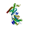 3t15C C: citing same article ( M: map data used to model this data |
|---|---|
| Similar structure data |
-Links
 PDBj
PDBj- Assembly
| Deposited unit | 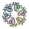
|
|---|---|
| 1 |
|
-Components
| #1: Protein | Mass: 32856.801 Da / Num. of mol.: 6 / Fragment: AAA ATPASE DOMAIN, RESIDUES 127-419 Source method: isolated from a genetically manipulated source Source: (gene. exp.)  |
|---|
-Experimental details
-Experiment
| Experiment | Method: ELECTRON MICROSCOPY |
|---|---|
| EM experiment | Aggregation state: PARTICLE / 3D reconstruction method: single particle reconstruction |
- Sample preparation
| Component | Name: NICOTIANA TABACUM RUBISCO ACTIVASE (R294V) / Type: COMPLEX / Details: THE PROTEIN FOR EM CARRIES A R294V MUTATION |
|---|---|
| Buffer solution | Name: 20MM TRIS-HCL, 50MM NACL, 2MM MGCL2,1MM ATPGAMMAS / pH: 8 / Details: 20MM TRIS-HCL, 50MM NACL, 2MM MGCL2,1MM ATPGAMMAS |
| Specimen | Conc.: 0.04 mg/ml / Embedding applied: NO / Shadowing applied: NO / Staining applied: YES / Vitrification applied: NO |
| EM staining | Type: NEGATIVE / Material: uranyl acetate |
| Specimen support | Details: CARBON |
- Electron microscopy imaging
| Microscopy | Model: FEI TECNAI 12 / Date: Jun 22, 2011 |
|---|---|
| Electron gun | Electron source: LAB6 / Accelerating voltage: 120 kV / Illumination mode: FLOOD BEAM |
| Electron lens | Mode: BRIGHT FIELD / Nominal defocus max: 950 nm / Nominal defocus min: 470 nm / Cs: 2 mm |
| Image recording | Electron dose: 20 e/Å2 / Film or detector model: FEI EAGLE (2k x 2k) |
| Image scans | Num. digital images: 11 |
| Radiation wavelength | Relative weight: 1 |
- Processing
| EM software |
| |||||||||||||||
|---|---|---|---|---|---|---|---|---|---|---|---|---|---|---|---|---|
| CTF correction | Details: PHASE FLIPPING, EACH PARTICLE | |||||||||||||||
| Symmetry | Point symmetry: C6 (6 fold cyclic) | |||||||||||||||
| 3D reconstruction | Method: ANGULAR RECONSTITUTION / Resolution: 20 Å / Num. of particles: 599 / Nominal pixel size: 3.308 Å / Actual pixel size: 3.308 Å Details: A MODULE OF ALPHA HELICAL DOMAIN AND ALPHA-BETA DOMAIN OF THE NEIGHBOURING SUBUNIT WAS OVERLAID WITH THE P97 D2 (3CF3) STRUCTURE AND THE HEXAMER WAS FITTED INTO THE EM MAP SUBMISSION BASED ...Details: A MODULE OF ALPHA HELICAL DOMAIN AND ALPHA-BETA DOMAIN OF THE NEIGHBOURING SUBUNIT WAS OVERLAID WITH THE P97 D2 (3CF3) STRUCTURE AND THE HEXAMER WAS FITTED INTO THE EM MAP SUBMISSION BASED ON EXPERIMENTAL DATA FROM EMDB EMD-1940. (DEPOSITION ID: 10169). Symmetry type: POINT | |||||||||||||||
| Atomic model building | Protocol: RIGID BODY FIT / Space: REAL / Target criteria: LOCAL MINIMISATION, FIT IN MAP / Details: METHOD--RIGID BODY REFINEMENT PROTOCOL--X-RAY | |||||||||||||||
| Atomic model building | PDB-ID: 3T15 Accession code: 3T15 / Source name: PDB / Type: experimental model | |||||||||||||||
| Refinement | Highest resolution: 20 Å | |||||||||||||||
| Refinement step | Cycle: LAST / Highest resolution: 20 Å
|
