- PDB-3syn: Crystal structure of FlhF in complex with its activator -
+
Open data
ID or keywords:
Loading...
-
Basic information
Entry
Database: PDB / ID: 3syn
Title
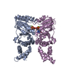
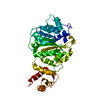
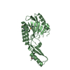
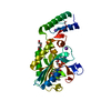
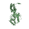
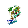
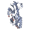
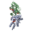
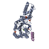
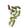
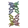
Crystal structure of FlhF in complex with its activator
Components
ATP-binding protein YlxH
Flagellar biosynthesis protein flhF
Keywords
PROTEIN TRANSPORT / SRP GTPASE / FLAGELLUM / BIOSYNTHETIC PROTEIN / GTPase ACTIVATING PROTEIN / Type 3 secretion system / GTP-binding protein
Function / homology
Function and homology information
bacterial-type flagellum organization / signal recognition particle binding / SRP-dependent cotranslational protein targeting to membrane / protein targeting / cytoplasmic side of plasma membrane / protein transport / GTPase activity / GTP binding / ATP hydrolysis activity / ATP binding ...bacterial-type flagellum organization / signal recognition particle binding / SRP-dependent cotranslational protein targeting to membrane / protein targeting / cytoplasmic side of plasma membrane / protein transport / GTPase activity / GTP binding / ATP hydrolysis activity / ATP binding / identical protein binding / plasma membrane / cytosol Similarity search - Function
Flagellar FlhF biosynthesis protein, N domain / : / Flagellar biosynthesis protein FlhF, N domain / Flagellar biosynthesis protein FlhF / : / Flagellum site-determining protein FlhG / Flagellum site-determining protein YlxH/ Fe-S cluster assembling factor NBP35 / NUBPL iron-transfer P-loop NTPase / ATP binding protein MinD/FleN / : ...Flagellar FlhF biosynthesis protein, N domain / : / Flagellar biosynthesis protein FlhF, N domain / Flagellar biosynthesis protein FlhF / : / Flagellum site-determining protein FlhG / Flagellum site-determining protein YlxH/ Fe-S cluster assembling factor NBP35 / NUBPL iron-transfer P-loop NTPase / ATP binding protein MinD/FleN / : / Signal recognition particle, SRP54 subunit, GTPase domain / SRP54-type protein, GTPase domain / SRP54-type protein, GTPase domain / Four Helix Bundle (Hemerythrin (Met), subunit A) / P-loop containing nucleotide triphosphate hydrolases / Up-down Bundle / Rossmann fold / P-loop containing nucleoside triphosphate hydrolase / 3-Layer(aba) Sandwich / Mainly Alpha / Alpha Beta Similarity search - Domain/homology
ALUMINUM FLUORIDE / GUANOSINE-5'-DIPHOSPHATE / Flagellum site-determining protein YlxH / Flagellar biosynthesis protein FlhF Similarity search - Component
A: Flagellar biosynthesis protein flhF B: Flagellar biosynthesis protein flhF C: Flagellar biosynthesis protein flhF D: Flagellar biosynthesis protein flhF E: ATP-binding protein YlxH F: ATP-binding protein YlxH G: ATP-binding protein YlxH H: ATP-binding protein YlxH hetero molecules
In the structure databanks used in Yorodumi, some data are registered as the other names, "COVID-19 virus" and "2019-nCoV". Here are the details of the virus and the list of structure data.
Jan 31, 2019. EMDB accession codes are about to change! (news from PDBe EMDB page)
EMDB accession codes are about to change! (news from PDBe EMDB page)
The allocation of 4 digits for EMDB accession codes will soon come to an end. Whilst these codes will remain in use, new EMDB accession codes will include an additional digit and will expand incrementally as the available range of codes is exhausted. The current 4-digit format prefixed with “EMD-” (i.e. EMD-XXXX) will advance to a 5-digit format (i.e. EMD-XXXXX), and so on. It is currently estimated that the 4-digit codes will be depleted around Spring 2019, at which point the 5-digit format will come into force.
The EM Navigator/Yorodumi systems omit the EMD- prefix.
Related info.:Q: What is EMD? / ID/Accession-code notation in Yorodumi/EM Navigator
Yorodumi is a browser for structure data from EMDB, PDB, SASBDB, etc.
This page is also the successor to EM Navigator detail page, and also detail information page/front-end page for Omokage search.
The word "yorodu" (or yorozu) is an old Japanese word meaning "ten thousand". "mi" (miru) is to see.
Related info.:EMDB / PDB / SASBDB / Comparison of 3 databanks / Yorodumi Search / Aug 31, 2016. New EM Navigator & Yorodumi / Yorodumi Papers / Jmol/JSmol / Function and homology information / Changes in new EM Navigator and Yorodumi
 Movie
Movie Controller


 Open data
 Basic information
 Components
 Keywords
 Function and homology information
 X-RAY DIFFRACTION /
  Authors
 Citation
 Structure visualization
 Downloads & links
 Other downloads
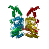


 PDBj
PDBj

 Assembly


 Type: RNA linking / Mass: 443.201 Da / Num. of mol.: 4 / Source method: obtained synthetically / Formula: C10H15N5O11P2 / Comment: GDP, energy-carrying molecule*YM
 Mass: 24.305 Da / Num. of mol.: 4 / Source method: obtained synthetically / Formula: Mg
 Mass: 83.977 Da / Num. of mol.: 4 / Source method: obtained synthetically / Formula: AlF3
 Mass: 96.063 Da / Num. of mol.: 15 / Source method: obtained synthetically / Formula: SO4
 Sample preparation
 / Beamline: ID14-4 / Wavelength: 1 Å
 Processing