 Movie
Movie Controller

[English] 日本語
 Yorodumi
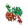
- PDB-6aan: Crystal structure of Methanosarcina mazei PylRS(Y306A/Y384F) comp... -
+ Open data

- Basic information
| Entry | Database: PDB / ID: 6aan | ||||||
|---|---|---|---|---|---|---|---|
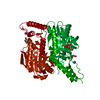
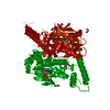
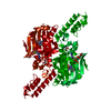
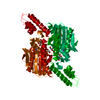
| Title | Crystal structure of Methanosarcina mazei PylRS(Y306A/Y384F) complexed with mEtZLys | ||||||
 Components Components | Pyrrolysine--tRNA ligase | ||||||
 Keywords Keywords | TRANSLATION / aminoacyl-tRNA synthetase / non-natural amino acids | ||||||
| Function / homology |  Function and homology information Function and homology informationpyrrolysine-tRNAPyl ligase / pyrrolysyl-tRNA synthetase activity / tRNA aminoacylation for protein translation / tRNA binding / ATP binding / cytoplasm Similarity search - Function | ||||||
| Biological species |  Methanosarcina mazei JCM 9314 (archaea) Methanosarcina mazei JCM 9314 (archaea) | ||||||
| Method |  X-RAY DIFFRACTION / SYNCHROTRON / MOLECULAR REPLACEMENT / Resolution: 1.51 Å X-RAY DIFFRACTION / SYNCHROTRON / MOLECULAR REPLACEMENT / Resolution: 1.51 Å | ||||||
 Authors Authors | Yanagisawa, T. / Kuratani, M. / Yokoyama, S. | ||||||
| Funding support |  Japan, 1items Japan, 1items
| ||||||
 Citation Citation | Journal: Cell Chem Biol / Year: 2019 Title: Structural Basis for Genetic-Code Expansion with Bulky Lysine Derivatives by an Engineered Pyrrolysyl-tRNA Synthetase. Authors: Yanagisawa, T. / Kuratani, M. / Seki, E. / Hino, N. / Sakamoto, K. / Yokoyama, S. | ||||||
| History |
|
- Structure visualization
| Structure viewer | Molecule: MolmilJmol/JSmol |
|---|
- Downloads & links
-Download
| PDBx/mmCIF format | 6aan.cif.gz | 82.5 KB | Display | PDBx/mmCIF format |
|---|---|---|---|---|
| PDB format | pdb6aan.ent.gz | 57.8 KB | Display | PDB format |
| PDBx/mmJSON format | 6aan.json.gz | Tree view | PDBx/mmJSON format | |
| Others |  Other downloads Other downloads |
-Validation report
| Arichive directory | https://data.pdbj.org/pub/pdb/validation_reports/aa/6aanftp://data.pdbj.org/pub/pdb/validation_reports/aa/6aan | HTTPS FTP |
|---|
-Related structure data
| Related structure data |  6aacC  6aadC  6aaoC 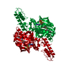 6aapC 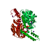 6aaqC  6aazC 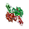 6ab0C 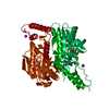 6ab1C 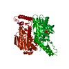 6ab2C 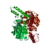 6ab8C 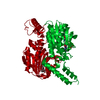 6abkC 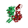 6ablC 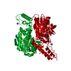 6abmC 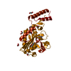 2zimS C: citing same article ( S: Starting model for refinement |
|---|---|
| Similar structure data |


-Links
 PDBj
PDBj
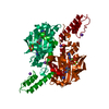
- Assembly
| Deposited unit | 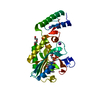
| ||||||||
|---|---|---|---|---|---|---|---|---|---|
| 1 | 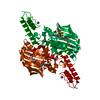
| ||||||||
| Unit cell |
|
-Components
-Protein , 1 types, 1 molecules A
| #1: Protein | Mass: 31346.982 Da / Num. of mol.: 1 / Mutation: Y306A,Y384F Source method: isolated from a genetically manipulated source Source: (gene. exp.) Methanosarcina mazei JCM 9314 (archaea)Gene: pylS / Production host:  |
|---|
-Non-polymers , 6 types, 237 molecules 


| #2: Chemical |  Mass: 24.305 Da / Num. of mol.: 3 / Source method: obtained synthetically / Formula: Mg Mass: 24.305 Da / Num. of mol.: 3 / Source method: obtained synthetically / Formula: Mg#3: Chemical | ChemComp-ATP / |  Mass: 507.181 Da / Num. of mol.: 1 / Source method: obtained synthetically / Formula: C10H16N5O13P3 / Comment: ATP, energy-carrying molecule*YM Mass: 507.181 Da / Num. of mol.: 1 / Source method: obtained synthetically / Formula: C10H16N5O13P3 / Comment: ATP, energy-carrying molecule*YM#4: Chemical | ChemComp-9TX / |  Type: peptide linking / Mass: 304.341 Da / Num. of mol.: 1 / Source method: obtained synthetically / Formula: C16H20N2O4 Type: peptide linking / Mass: 304.341 Da / Num. of mol.: 1 / Source method: obtained synthetically / Formula: C16H20N2O4#5: Chemical |  Mass: 39.098 Da / Num. of mol.: 2 / Source method: obtained synthetically / Formula: K Mass: 39.098 Da / Num. of mol.: 2 / Source method: obtained synthetically / Formula: K#6: Chemical | ChemComp-PEG / |  Mass: 106.120 Da / Num. of mol.: 1 / Source method: obtained synthetically / Formula: C4H10O3 Mass: 106.120 Da / Num. of mol.: 1 / Source method: obtained synthetically / Formula: C4H10O3#7: Water | ChemComp-HOH / | Mass: 18.015 Da / Num. of mol.: 229 / Source method: isolated from a natural source / Formula: H2O |
|---|
-Details
| Sequence details | The sequence reference Q8PWY1 (PYLS_METMA) is derived from a different strain (Methanosarcina mazei ...The sequence reference Q8PWY1 (PYLS_METMA) is derived from a different strain (Methanosarcina mazei Go1, TaxID 192952). Residue E444G is a natural mutation observed in PylRS gene from Methanosarcina mazei JCM 9314 genome. |
|---|
-Experimental details
-Experiment
| Experiment | Method: X-RAY DIFFRACTION / Number of used crystals: 1 |
|---|
- Sample preparation
| Crystal | Density Matthews: 2.27 Å3/Da / Density % sol: 45.88 % |
|---|---|
| Crystal grow | Temperature: 281 K / Method: vapor diffusion, hanging drop / pH: 8.5 Details: Tris-HCl (pH 8.5), PEG 200, KCl, MgCl2, sodium citrate |
-Data collection
| Diffraction | Mean temperature: 100 K |
|---|---|
| Diffraction source | Source: SYNCHROTRON / Site: SPring-8 / Beamline: BL32XU / Wavelength: 1 Å |
| Detector | Type: RAYONIX MX225-HS / Detector: CCD / Date: Apr 21, 2015 |
| Radiation | Protocol: SINGLE WAVELENGTH / Monochromatic (M) / Laue (L): M / Scattering type: x-ray |
| Radiation wavelength | Wavelength: 1 Å / Relative weight: 1 |
| Reflection | Resolution: 1.51→50 Å / Num. obs: 43126 / % possible obs: 97.3 % / Redundancy: 3.8 % / Rpim(I) all: 0.029 / Rrim(I) all: 0.057 / Net I/σ(I): 36.5 |
| Reflection shell | Resolution: 1.51→1.55 Å / Num. unique obs: 2133 / Rpim(I) all: 0.291 |
- Processing
| Software |
| |||||||||||||||||||||||||||||||||||||||||||||||||||||||||||||||||||||||||||||||||||||||||||||||||||||||||
|---|---|---|---|---|---|---|---|---|---|---|---|---|---|---|---|---|---|---|---|---|---|---|---|---|---|---|---|---|---|---|---|---|---|---|---|---|---|---|---|---|---|---|---|---|---|---|---|---|---|---|---|---|---|---|---|---|---|---|---|---|---|---|---|---|---|---|---|---|---|---|---|---|---|---|---|---|---|---|---|---|---|---|---|---|---|---|---|---|---|---|---|---|---|---|---|---|---|---|---|---|---|---|---|---|---|---|
| Refinement | Method to determine structure: MOLECULAR REPLACEMENT Starting model: 2zim Resolution: 1.51→35.004 Å / SU ML: 0.14 / Cross valid method: NONE / σ(F): 1.36 / Phase error: 21.4
| |||||||||||||||||||||||||||||||||||||||||||||||||||||||||||||||||||||||||||||||||||||||||||||||||||||||||
| Solvent computation | Shrinkage radii: 0.9 Å / VDW probe radii: 1.11 Å | |||||||||||||||||||||||||||||||||||||||||||||||||||||||||||||||||||||||||||||||||||||||||||||||||||||||||
| Refinement step | Cycle: LAST / Resolution: 1.51→35.004 Å
| |||||||||||||||||||||||||||||||||||||||||||||||||||||||||||||||||||||||||||||||||||||||||||||||||||||||||
| Refine LS restraints |
| |||||||||||||||||||||||||||||||||||||||||||||||||||||||||||||||||||||||||||||||||||||||||||||||||||||||||
| LS refinement shell |
|
