 Movie
Movie Controller

[English] 日本語
 Yorodumi
- PDB-3syi: Crystal structure of sulfide:quinone oxidoreductase Ser126Ala var... -
+ Open data

- Basic information
| Entry | Database: PDB / ID: 3syi | ||||||
|---|---|---|---|---|---|---|---|
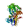
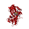
| Title | Crystal structure of sulfide:quinone oxidoreductase Ser126Ala variant from Acidithiobacillus ferrooxidans using 7.0 keV diffraction data | ||||||
 Components Components | Sulfide-quinone reductase, putative | ||||||
 Keywords Keywords | OXIDOREDUCTASE / sulfide:quinone oxidoreductase / Ser126Ala variant / integral monotopic membrane protein / complex with sulfide | ||||||
| Function / homology |  Function and homology information Function and homology informationbacterial sulfide:quinone reductase / sulfide:quinone oxidoreductase activity / NAD(P)H dehydrogenase (quinone) activity / aerobic electron transport chain / quinone binding / nucleotide binding / membrane Similarity search - Function | ||||||
| Biological species |  Acidithiobacillus ferrooxidans (bacteria) Acidithiobacillus ferrooxidans (bacteria) | ||||||
| Method |  X-RAY DIFFRACTION / SYNCHROTRON / refinement / Resolution: 2.2001 Å X-RAY DIFFRACTION / SYNCHROTRON / refinement / Resolution: 2.2001 Å | ||||||
 Authors Authors | Cherney, M.M. / Zhang, Y. / James, M.N.G. / Weiner, J.H. | ||||||
 Citation Citation | Journal: J.Struct.Biol. / Year: 2012 Title: Structure-activity characterization of sulfide:quinone oxidoreductase variants. Authors: Cherney, M.M. / Zhang, Y. / James, M.N. / Weiner, J.H. | ||||||
| History |
|
- Structure visualization
| Structure viewer | Molecule: MolmilJmol/JSmol |
|---|
- Downloads & links
-Download
| PDBx/mmCIF format | 3syi.cif.gz | 102.4 KB | Display | PDBx/mmCIF format |
|---|---|---|---|---|
| PDB format | pdb3syi.ent.gz | 75.7 KB | Display | PDB format |
| PDBx/mmJSON format | 3syi.json.gz | Tree view | PDBx/mmJSON format | |
| Others |  Other downloads Other downloads |
-Validation report
| Arichive directory | https://data.pdbj.org/pub/pdb/validation_reports/sy/3syiftp://data.pdbj.org/pub/pdb/validation_reports/sy/3syi | HTTPS FTP |
|---|
-Related structure data
| Related structure data | 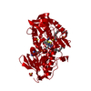 3sx6C 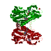 3sxiC  3sy4C  3sz0C 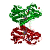 3szcC 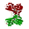 3szfC 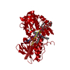 3szwC  3t0kC  3t14C  3t2kC  3t2yC  3kpg C: citing same article ( S: Starting model for refinement |
|---|---|
| Similar structure data |


-Links
 PDBj
PDBj


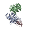
- Assembly
| Deposited unit | 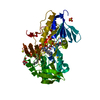
| ||||||||
|---|---|---|---|---|---|---|---|---|---|
| 1 | 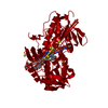
| ||||||||
| Unit cell |
|
-Components
-Protein / Sugars , 2 types, 2 molecules A
| #1: Protein | Mass: 47749.957 Da / Num. of mol.: 1 / Mutation: S126A Source method: isolated from a genetically manipulated source Source: (gene. exp.) Acidithiobacillus ferrooxidans (bacteria)Strain: ATCC 23270 / DSM 14882 / NCIB 8455 / Gene: AFE_1792 / Production host: |
|---|---|
| #3: Sugar | ChemComp-LMT /  Type: D-saccharide / Mass: 510.615 Da / Num. of mol.: 1 / Source method: obtained synthetically / Formula: C24H46O11 / Comment: detergent*YM Type: D-saccharide / Mass: 510.615 Da / Num. of mol.: 1 / Source method: obtained synthetically / Formula: C24H46O11 / Comment: detergent*YM |
-Non-polymers , 4 types, 111 molecules 


| #2: Chemical | ChemComp-FAD /  Mass: 785.550 Da / Num. of mol.: 1 / Source method: obtained synthetically / Formula: C27H33N9O15P2 / Comment: FAD*YM Mass: 785.550 Da / Num. of mol.: 1 / Source method: obtained synthetically / Formula: C27H33N9O15P2 / Comment: FAD*YM | ||||
|---|---|---|---|---|---|
| #4: Chemical | ChemComp-H2S /  Mass: 34.081 Da / Num. of mol.: 5 / Source method: obtained synthetically / Formula: H2S Mass: 34.081 Da / Num. of mol.: 5 / Source method: obtained synthetically / Formula: H2S#5: Chemical | ChemComp-SO4 / |  Mass: 96.063 Da / Num. of mol.: 1 / Source method: obtained synthetically / Formula: SO4 Mass: 96.063 Da / Num. of mol.: 1 / Source method: obtained synthetically / Formula: SO4#6: Water | ChemComp-HOH / | Mass: 18.015 Da / Num. of mol.: 104 / Source method: isolated from a natural source / Formula: H2O |
-Experimental details
-Experiment
| Experiment | Method: X-RAY DIFFRACTION / Number of used crystals: 1 |
|---|
- Sample preparation
| Crystal | Density Matthews: 2.78 Å3/Da / Density % sol: 55.72 % |
|---|---|
| Crystal grow | Temperature: 295 K / Method: vapor diffusion, hanging drop / pH: 5.5 Details: 30% PEG 600, 0.1 M bis-tris buffer,0.1 M MgSO4, 0.05% DDM, pH 5.5, VAPOR DIFFUSION, HANGING DROP, temperature 295K |
-Data collection
| Diffraction | Mean temperature: 100 K |
|---|---|
| Diffraction source | Source: SYNCHROTRON / Site: CLSI  / Beamline: 08ID-1 / Wavelength: 1.7712 Å / Beamline: 08ID-1 / Wavelength: 1.7712 Å |
| Detector | Type: MARMOSAIC 300 mm CCD / Detector: CCD / Date: Feb 12, 2010 |
| Radiation | Monochromator: ACCEL/BRUKER double crystal monochromator (DCM), featuring indirectly cryo-cooled first crystal and sagittally focusing second crystal Protocol: SINGLE WAVELENGTH / Monochromatic (M) / Laue (L): M / Scattering type: x-ray |
| Radiation wavelength | Wavelength: 1.7712 Å / Relative weight: 1 |
| Reflection | Resolution: 2.2→49 Å / Num. obs: 48437 / % possible obs: 94.5 % / Observed criterion σ(F): 0 / Observed criterion σ(I): 0 / Redundancy: 12.2 % / Rmerge(I) obs: 0.098 / Rsym value: 0.098 / Net I/σ(I): 14.5 |
| Reflection shell | Resolution: 2.2→2.32 Å / Redundancy: 2.6 % / Rmerge(I) obs: 0.833 / Mean I/σ(I) obs: 1.3 / Rsym value: 0.833 / % possible all: 69.2 |
- Processing
| Software |
| ||||||||||||||||||||||||||||||||||||||||||||||||||||||||||||||||||||||||||||||||||||||||||||||||||||||||||||||||||||||||||||||
|---|---|---|---|---|---|---|---|---|---|---|---|---|---|---|---|---|---|---|---|---|---|---|---|---|---|---|---|---|---|---|---|---|---|---|---|---|---|---|---|---|---|---|---|---|---|---|---|---|---|---|---|---|---|---|---|---|---|---|---|---|---|---|---|---|---|---|---|---|---|---|---|---|---|---|---|---|---|---|---|---|---|---|---|---|---|---|---|---|---|---|---|---|---|---|---|---|---|---|---|---|---|---|---|---|---|---|---|---|---|---|---|---|---|---|---|---|---|---|---|---|---|---|---|---|---|---|---|
| Refinement | Method to determine structure: refinement Starting model: PDB ENTRY 3KPG 3kpg Resolution: 2.2001→42.069 Å / SU ML: 0.7 / σ(F): 1.34 / Phase error: 27.27 / Stereochemistry target values: ML
| ||||||||||||||||||||||||||||||||||||||||||||||||||||||||||||||||||||||||||||||||||||||||||||||||||||||||||||||||||||||||||||||
| Solvent computation | Shrinkage radii: 0.83 Å / VDW probe radii: 1.1 Å / Solvent model: FLAT BULK SOLVENT MODEL / Bsol: 39.876 Å2 / ksol: 0.332 e/Å3 | ||||||||||||||||||||||||||||||||||||||||||||||||||||||||||||||||||||||||||||||||||||||||||||||||||||||||||||||||||||||||||||||
| Displacement parameters |
| ||||||||||||||||||||||||||||||||||||||||||||||||||||||||||||||||||||||||||||||||||||||||||||||||||||||||||||||||||||||||||||||
| Refinement step | Cycle: LAST / Resolution: 2.2001→42.069 Å
| ||||||||||||||||||||||||||||||||||||||||||||||||||||||||||||||||||||||||||||||||||||||||||||||||||||||||||||||||||||||||||||||
| Refine LS restraints |
| ||||||||||||||||||||||||||||||||||||||||||||||||||||||||||||||||||||||||||||||||||||||||||||||||||||||||||||||||||||||||||||||
| LS refinement shell |
|
