 Movie
Movie Controller

[English] 日本語
 Yorodumi
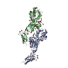
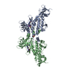
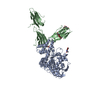
- PDB-3sy2: Crystal structure of the Salmonella E3 ubiquitin ligase SopA in c... -
+ Open data

- Basic information
| Entry | Database: PDB / ID: 3sy2 | ||||||
|---|---|---|---|---|---|---|---|
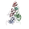
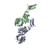
| Title | Crystal structure of the Salmonella E3 ubiquitin ligase SopA in complex with the human E2 UbcH7 | ||||||
 Components Components |
| ||||||
 Keywords Keywords | LIGASE/SIGNALING PROTEIN / pentapeptide / HECT domain / HECT E3 / HECT E3 ubiquitin ligase / E2 ubiquitin conjugating enzyme / ubiquitin / protein-protein complex / effector protein / ubiquitin transfer / ubiquitination / LIGASE-SIGNALING PROTEIN complex | ||||||
| Function / homology |  Function and homology information Function and homology informationcell cycle phase transition / ubiquitin-protein transferase activator activity / HECT-type E3 ubiquitin transferase / protein K11-linked ubiquitination / : / cellular response to glucocorticoid stimulus / E2 ubiquitin-conjugating enzyme / cellular response to steroid hormone stimulus / ubiquitin conjugating enzyme activity / ubiquitin ligase complex ...cell cycle phase transition / ubiquitin-protein transferase activator activity / HECT-type E3 ubiquitin transferase / protein K11-linked ubiquitination / : / cellular response to glucocorticoid stimulus / E2 ubiquitin-conjugating enzyme / cellular response to steroid hormone stimulus / ubiquitin conjugating enzyme activity / ubiquitin ligase complex / protein modification process / positive regulation of protein ubiquitination / Synthesis of active ubiquitin: roles of E1 and E2 enzymes / PINK1-PRKN Mediated Mitophagy / Regulation of TNFR1 signaling / Regulation of necroptotic cell death / protein polyubiquitination / ubiquitin-protein transferase activity / ubiquitin protein ligase activity / host cell / Antigen processing: Ubiquitination & Proteasome degradation / E3 ubiquitin ligases ubiquitinate target proteins / ubiquitin-dependent protein catabolic process / transcription coactivator activity / cell population proliferation / protein ubiquitination / ubiquitin protein ligase binding / regulation of DNA-templated transcription / enzyme binding / RNA binding / extracellular region / nucleoplasm / ATP binding / nucleus / cytoplasm / cytosol Similarity search - Function | ||||||
| Biological species |  Salmonella enterica subsp. enterica serovar Typhimurium (bacteria) Salmonella enterica subsp. enterica serovar Typhimurium (bacteria) Homo sapiens (human) Homo sapiens (human) | ||||||
| Method |  X-RAY DIFFRACTION / SYNCHROTRON / MOLECULAR REPLACEMENT / Resolution: 3.27 Å X-RAY DIFFRACTION / SYNCHROTRON / MOLECULAR REPLACEMENT / Resolution: 3.27 Å | ||||||
 Authors Authors | Diao, J. / Lin, D.Y. / Chen, J. | ||||||
 Citation Citation | Journal: Proc.Natl.Acad.Sci.USA / Year: 2012 Title: Crystal structures of two bacterial HECT-like E3 ligases in complex with a human E2 reveal atomic details of pathogen-host interactions. Authors: Lin, D.Y. / Diao, J. / Chen, J. | ||||||
| History |
|
- Structure visualization
| Structure viewer | Molecule: MolmilJmol/JSmol |
|---|
- Downloads & links
-Download
| PDBx/mmCIF format | 3sy2.cif.gz | 585.9 KB | Display | PDBx/mmCIF format |
|---|---|---|---|---|
| PDB format | pdb3sy2.ent.gz | 485.9 KB | Display | PDB format |
| PDBx/mmJSON format | 3sy2.json.gz | Tree view | PDBx/mmJSON format | |
| Others |  Other downloads Other downloads |
-Validation report
| Arichive directory | https://data.pdbj.org/pub/pdb/validation_reports/sy/3sy2ftp://data.pdbj.org/pub/pdb/validation_reports/sy/3sy2 | HTTPS FTP |
|---|
-Related structure data
| Related structure data |  3sqvC 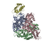 1c4zS 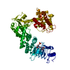 2qyuS S: Starting model for refinement C: citing same article ( |
|---|---|
| Similar structure data |


-Links
 PDBj
PDBj


- Assembly
| Deposited unit | 
| ||||||||
|---|---|---|---|---|---|---|---|---|---|
| 1 | 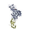
| ||||||||
| 2 | 
| ||||||||
| Unit cell |
|
-Components
| #1: Protein | Mass: 69235.219 Da / Num. of mol.: 2 Fragment: C-terminal beta-helix domain and HECT-like domain (UNP residues 165-782) Source method: isolated from a genetically manipulated source Source: (gene. exp.) Salmonella enterica subsp. enterica serovar Typhimurium (bacteria)Gene: sopA, STM2066 / Plasmid: pMCSG7 / Production host: References: UniProt: Q8ZNR3, Ligases; Forming carbon-nitrogen bonds; Acid-amino-acid ligases (peptide synthases) #2: Protein | Mass: 18033.723 Da / Num. of mol.: 2 Source method: isolated from a genetically manipulated source Source: (gene. exp.) Homo sapiens (human) / Gene: UBE2L3, UBCE7, UBCH7 / Plasmid: pGEX / Production host: #3: Chemical | ChemComp-SO4 / |   Mass: 96.063 Da / Num. of mol.: 1 / Source method: obtained synthetically / Formula: SO4 Mass: 96.063 Da / Num. of mol.: 1 / Source method: obtained synthetically / Formula: SO4#4: Water | ChemComp-HOH / |  Mass: 18.015 Da / Num. of mol.: 15 / Source method: isolated from a natural source / Formula: H2O Mass: 18.015 Da / Num. of mol.: 15 / Source method: isolated from a natural source / Formula: H2O |
|---|
-Experimental details
-Experiment
| Experiment | Method: X-RAY DIFFRACTION / Number of used crystals: 1 |
|---|
- Sample preparation
| Crystal | Density Matthews: 3.04 Å3/Da / Density % sol: 59.5 % |
|---|---|
| Crystal grow | Temperature: 298 K / Method: vapor diffusion, sitting drop / pH: 6.5 Details: 0.1 M MES, 1.6 M ammonium sulfate, pH 6.5, VAPOR DIFFUSION, SITTING DROP, temperature 298K |
-Data collection
| Diffraction | Mean temperature: 100 K |
|---|---|
| Diffraction source | Source: SYNCHROTRON / Site: APS  / Beamline: 23-ID-D / Wavelength: 1.03324 Å / Beamline: 23-ID-D / Wavelength: 1.03324 Å |
| Detector | Type: MARMOSAIC 300 mm CCD / Detector: CCD / Date: Jul 26, 2008 |
| Radiation | Monochromator: Si(111) / Protocol: SINGLE WAVELENGTH / Monochromatic (M) / Laue (L): M / Scattering type: x-ray |
| Radiation wavelength | Wavelength: 1.03324 Å / Relative weight: 1 |
| Reflection | Resolution: 3.2→50 Å / Num. all: 31621 / Num. obs: 31621 / % possible obs: 86.7 % / Observed criterion σ(F): 0 / Observed criterion σ(I): 0 / Redundancy: 3.1 % / Biso Wilson estimate: 85.18 Å2 / Rsym value: 0.147 / Net I/σ(I): 7.19 |
| Reflection shell | Resolution: 3.2→3.31 Å / Redundancy: 2.7 % / Rmerge(I) obs: 0.52 / Mean I/σ(I) obs: 2.39 / Num. unique all: 3105 / Rsym value: 0.52 / % possible all: 83.7 |
- Processing
| Software |
| |||||||||||||||||||||||||||||||||||||||||||||||||||||||||||||||||||||||||||||||||||||||||||||||||||||||||||||||||||||||||||||
|---|---|---|---|---|---|---|---|---|---|---|---|---|---|---|---|---|---|---|---|---|---|---|---|---|---|---|---|---|---|---|---|---|---|---|---|---|---|---|---|---|---|---|---|---|---|---|---|---|---|---|---|---|---|---|---|---|---|---|---|---|---|---|---|---|---|---|---|---|---|---|---|---|---|---|---|---|---|---|---|---|---|---|---|---|---|---|---|---|---|---|---|---|---|---|---|---|---|---|---|---|---|---|---|---|---|---|---|---|---|---|---|---|---|---|---|---|---|---|---|---|---|---|---|---|---|---|
| Refinement | Method to determine structure: MOLECULAR REPLACEMENT Starting model: PDB ENTRIES 2QYU AND 1C4Z Resolution: 3.27→45.424 Å / SU ML: 0.42 / σ(F): 1.34 / Phase error: 27.69 / Stereochemistry target values: ML
| |||||||||||||||||||||||||||||||||||||||||||||||||||||||||||||||||||||||||||||||||||||||||||||||||||||||||||||||||||||||||||||
| Solvent computation | Shrinkage radii: 0.9 Å / VDW probe radii: 1.11 Å / Solvent model: FLAT BULK SOLVENT MODEL / Bsol: 55.369 Å2 / ksol: 0.307 e/Å3 | |||||||||||||||||||||||||||||||||||||||||||||||||||||||||||||||||||||||||||||||||||||||||||||||||||||||||||||||||||||||||||||
| Displacement parameters |
| |||||||||||||||||||||||||||||||||||||||||||||||||||||||||||||||||||||||||||||||||||||||||||||||||||||||||||||||||||||||||||||
| Refinement step | Cycle: LAST / Resolution: 3.27→45.424 Å
| |||||||||||||||||||||||||||||||||||||||||||||||||||||||||||||||||||||||||||||||||||||||||||||||||||||||||||||||||||||||||||||
| Refine LS restraints |
| |||||||||||||||||||||||||||||||||||||||||||||||||||||||||||||||||||||||||||||||||||||||||||||||||||||||||||||||||||||||||||||
| LS refinement shell |
| |||||||||||||||||||||||||||||||||||||||||||||||||||||||||||||||||||||||||||||||||||||||||||||||||||||||||||||||||||||||||||||
| Refinement TLS params. | Method: refined / Refine-ID: X-RAY DIFFRACTION
| |||||||||||||||||||||||||||||||||||||||||||||||||||||||||||||||||||||||||||||||||||||||||||||||||||||||||||||||||||||||||||||
| Refinement TLS group |
|
