 Movie
Movie Controller

[English] 日本語
 Yorodumi
- PDB-3sus: Crystal structure of beta-hexosaminidase from Paenibacillus sp. T... -
+ Open data

- Basic information
| Entry | Database: PDB / ID: 3sus | ||||||
|---|---|---|---|---|---|---|---|
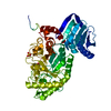
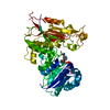
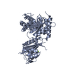
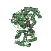
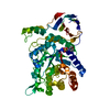
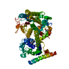
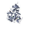
| Title | Crystal structure of beta-hexosaminidase from Paenibacillus sp. TS12 in complex with Gal-NAG-thiazoline | ||||||
 Components Components | Beta-hexosaminidase | ||||||
 Keywords Keywords | HYDROLASE / Structural Genomics / RIKEN Structural Genomics/Proteomics Initiative / RSGI / Tim Barrel / Carbohydrate/Sugar Binding | ||||||
| Function / homology |  Function and homology information Function and homology informationglycosaminoglycan metabolic process / beta-N-acetylhexosaminidase activity / beta-N-acetylhexosaminidase / polysaccharide catabolic process / carbohydrate binding / carbohydrate metabolic process / membrane Similarity search - Function | ||||||
| Biological species |  Paenibacillus (bacteria) Paenibacillus (bacteria) | ||||||
| Method |  X-RAY DIFFRACTION / SYNCHROTRON / MOLECULAR REPLACEMENT / Resolution: 1.8 Å X-RAY DIFFRACTION / SYNCHROTRON / MOLECULAR REPLACEMENT / Resolution: 1.8 Å | ||||||
 Authors Authors | Sumida, T. / Yokoyama, S. / RIKEN Structural Genomics/Proteomics Initiative (RSGI) | ||||||
 Citation Citation | Journal: Org.Biomol.Chem. / Year: 2012 Title: Gaining insight into the inhibition of glycoside hydrolase family 20 exo-beta-N-acetylhexosaminidases using a structural approach Authors: Sumida, T. / Stubbs, K.A. / Ito, M. / Yokoyama, S. | ||||||
| History |
|
- Structure visualization
| Structure viewer | Molecule: MolmilJmol/JSmol |
|---|
- Downloads & links
-Download
| PDBx/mmCIF format | 3sus.cif.gz | 225.8 KB | Display | PDBx/mmCIF format |
|---|---|---|---|---|
| PDB format | pdb3sus.ent.gz | 178.4 KB | Display | PDB format |
| PDBx/mmJSON format | 3sus.json.gz | Tree view | PDBx/mmJSON format | |
| Others |  Other downloads Other downloads |
-Validation report
| Summary document | 3sus_validation.pdf.gz | 458.8 KB | Display | wwPDB validaton report |
|---|---|---|---|---|
| Full document | 3sus_full_validation.pdf.gz | 461.5 KB | Display | |
| Data in XML | 3sus_validation.xml.gz | 25.4 KB | Display | |
| Data in CIF | 3sus_validation.cif.gz | 40.5 KB | Display | |
| Arichive directory | https://data.pdbj.org/pub/pdb/validation_reports/su/3susftp://data.pdbj.org/pub/pdb/validation_reports/su/3sus | HTTPS FTP |
-Related structure data
| Related structure data | 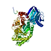 3surC  3sutC  3suuC 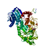 3suvC  3suwC 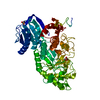 3gh4S C: citing same article ( S: Starting model for refinement |
|---|---|
| Similar structure data |


-Links
 PDBj
PDBj
- Assembly
| Deposited unit | 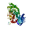
| ||||||||
|---|---|---|---|---|---|---|---|---|---|
| 1 |
| ||||||||
| Unit cell |
|
-Components
| #1: Protein | Mass: 57566.105 Da / Num. of mol.: 1 Source method: isolated from a genetically manipulated source Source: (gene. exp.) Paenibacillus (bacteria) / Strain: TS12 / Gene: Hex1 / Plasmid: pET28c / Production host: References: UniProt: D0VX21, UniProt: D2KW09*PLUS, beta-N-acetylhexosaminidase |
|---|---|
| #2: Chemical | ChemComp-SO4 /   Mass: 96.063 Da / Num. of mol.: 1 / Source method: obtained synthetically / Formula: SO4 Mass: 96.063 Da / Num. of mol.: 1 / Source method: obtained synthetically / Formula: SO4 |
| #3: Chemical | ChemComp-GNL / (  Mass: 219.258 Da / Num. of mol.: 1 / Source method: obtained synthetically / Formula: C8H13NO4S Mass: 219.258 Da / Num. of mol.: 1 / Source method: obtained synthetically / Formula: C8H13NO4S |
| #4: Water | ChemComp-HOH /  Mass: 18.015 Da / Num. of mol.: 644 / Source method: isolated from a natural source / Formula: H2O Mass: 18.015 Da / Num. of mol.: 644 / Source method: isolated from a natural source / Formula: H2O |
| Has protein modification | Y |
-Experimental details
-Experiment
| Experiment | Method: X-RAY DIFFRACTION / Number of used crystals: 1 |
|---|
- Sample preparation
| Crystal | Density Matthews: 2.28 Å3/Da / Density % sol: 46.06 % |
|---|---|
| Crystal grow | Temperature: 293 K / Method: vapor diffusion, hanging drop / pH: 4.6 Details: Na-citrate, PEG 2000, (NH4)2SO4, pH 4.6, VAPOR DIFFUSION, HANGING DROP, temperature 293K |
-Data collection
| Diffraction | Mean temperature: 100 K |
|---|---|
| Diffraction source | Source: SYNCHROTRON / Site: SPring-8  / Beamline: BL26B2 / Wavelength: 1 Å / Beamline: BL26B2 / Wavelength: 1 Å |
| Detector | Type: MARMOSAIC 225 mm CCD / Detector: CCD / Date: Apr 9, 2010 |
| Radiation | Protocol: SINGLE WAVELENGTH / Monochromatic (M) / Laue (L): M / Scattering type: x-ray |
| Radiation wavelength | Wavelength: 1 Å / Relative weight: 1 |
| Reflection | Resolution: 1.8→50 Å / Num. obs: 49006 / Redundancy: 7.3 % / Rsym value: 0.159 |
| Reflection shell | Resolution: 1.8→1.86 Å / Redundancy: 6.6 % / Rsym value: 0.521 |
- Processing
| Software |
| |||||||||||||||||||||||||||||||||||||||||||||||||||||||||||||||||||||||||||||||||||||||||||||||||||||||||
|---|---|---|---|---|---|---|---|---|---|---|---|---|---|---|---|---|---|---|---|---|---|---|---|---|---|---|---|---|---|---|---|---|---|---|---|---|---|---|---|---|---|---|---|---|---|---|---|---|---|---|---|---|---|---|---|---|---|---|---|---|---|---|---|---|---|---|---|---|---|---|---|---|---|---|---|---|---|---|---|---|---|---|---|---|---|---|---|---|---|---|---|---|---|---|---|---|---|---|---|---|---|---|---|---|---|---|
| Refinement | Method to determine structure: MOLECULAR REPLACEMENT Starting model: 3GH4 Resolution: 1.8→50 Å / Cor.coef. Fo:Fc: 0.963 / Cor.coef. Fo:Fc free: 0.94 / SU B: 4.73 / SU ML: 0.067 / Cross valid method: THROUGHOUT / σ(F): 0 / ESU R Free: 0.109 / Stereochemistry target values: MAXIMUM LIKELIHOOD / Details: HYDROGENS HAVE BEEN ADDED IN THE RIDING POSITIONS
| |||||||||||||||||||||||||||||||||||||||||||||||||||||||||||||||||||||||||||||||||||||||||||||||||||||||||
| Solvent computation | Ion probe radii: 0.8 Å / Shrinkage radii: 0.8 Å / VDW probe radii: 1.2 Å / Solvent model: MASK | |||||||||||||||||||||||||||||||||||||||||||||||||||||||||||||||||||||||||||||||||||||||||||||||||||||||||
| Displacement parameters | Biso mean: 12.868 Å2
| |||||||||||||||||||||||||||||||||||||||||||||||||||||||||||||||||||||||||||||||||||||||||||||||||||||||||
| Refinement step | Cycle: LAST / Resolution: 1.8→50 Å
| |||||||||||||||||||||||||||||||||||||||||||||||||||||||||||||||||||||||||||||||||||||||||||||||||||||||||
| Refine LS restraints |
| |||||||||||||||||||||||||||||||||||||||||||||||||||||||||||||||||||||||||||||||||||||||||||||||||||||||||
| LS refinement shell | Resolution: 1.8→1.847 Å / Total num. of bins used: 20
|
