 Movie
Movie Controller

[English] 日本語
 Yorodumi
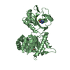
- PDB-3gh4: Crystal structure of beta-hexosaminidase from Paenibacillus sp. TS12 -
+ Open data

- Basic information
| Entry | Database: PDB / ID: 3gh4 | ||||||
|---|---|---|---|---|---|---|---|
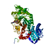
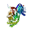
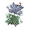
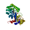
| Title | Crystal structure of beta-hexosaminidase from Paenibacillus sp. TS12 | ||||||
 Components Components | beta-hexosaminidase | ||||||
 Keywords Keywords | HYDROLASE / beta-N-acetylhexosaminidase / glycosphingolipids / Paenibacillus sp. / GH20 / Structural Genomics / NPPSFA / National Project on Protein Structural and Functional Analyses / RIKEN Structural Genomics/Proteomics Initiative / RSGI | ||||||
| Function / homology |  Function and homology information Function and homology informationglycosaminoglycan metabolic process / beta-N-acetylhexosaminidase activity / beta-N-acetylhexosaminidase / carbohydrate metabolic process / membrane Similarity search - Function | ||||||
| Biological species |  Paenibacillus sp. (bacteria) Paenibacillus sp. (bacteria) | ||||||
| Method |  X-RAY DIFFRACTION / SYNCHROTRON / MOLECULAR REPLACEMENT / Resolution: 1.8 Å X-RAY DIFFRACTION / SYNCHROTRON / MOLECULAR REPLACEMENT / Resolution: 1.8 Å | ||||||
 Authors Authors | Sumida, T. / Ishii, R. / Yanagisawa, T. / Yokoyama, S. / Ito, M. / RIKEN Structural Genomics/Proteomics Initiative (RSGI) | ||||||
 Citation Citation | Journal: J.Mol.Biol. / Year: 2009 Title: Molecular cloning and crystal structural analysis of a novel beta-N-acetylhexosaminidase from Paenibacillus sp. TS12 capable of degrading glycosphingolipids Authors: Sumida, T. / Ishii, R. / Yanagisawa, T. / Yokoyama, S. / Ito, M. | ||||||
| History |
|
- Structure visualization
| Structure viewer | Molecule: MolmilJmol/JSmol |
|---|
- Downloads & links
-Download
| PDBx/mmCIF format | 3gh4.cif.gz | 223.9 KB | Display | PDBx/mmCIF format |
|---|---|---|---|---|
| PDB format | pdb3gh4.ent.gz | 176.5 KB | Display | PDB format |
| PDBx/mmJSON format | 3gh4.json.gz | Tree view | PDBx/mmJSON format | |
| Others |  Other downloads Other downloads |
-Validation report
| Arichive directory | https://data.pdbj.org/pub/pdb/validation_reports/gh/3gh4ftp://data.pdbj.org/pub/pdb/validation_reports/gh/3gh4 | HTTPS FTP |
|---|
-Related structure data
| Related structure data |  3gh5C  3gh7C  1hp4S C: citing same article ( S: Starting model for refinement |
|---|---|
| Similar structure data |


-Links
 PDBj
PDBj


- Assembly
| Deposited unit | 
| ||||||||
|---|---|---|---|---|---|---|---|---|---|
| 1 |
| ||||||||
| Unit cell |
|
-Components
| #1: Protein | Mass: 57566.105 Da / Num. of mol.: 1 Source method: isolated from a genetically manipulated source Source: (gene. exp.) Paenibacillus sp. (bacteria) / Strain: TS12 / Gene: Hex1 / Plasmid: pET28c / Production host: References: UniProt: D0VX21*PLUS, beta-N-acetylhexosaminidase |
|---|---|
| #2: Chemical | ChemComp-SO4 /   Mass: 96.063 Da / Num. of mol.: 1 / Source method: obtained synthetically / Formula: SO4 Mass: 96.063 Da / Num. of mol.: 1 / Source method: obtained synthetically / Formula: SO4 |
| #3: Chemical | ChemComp-ACY /   Mass: 60.052 Da / Num. of mol.: 1 / Source method: obtained synthetically / Formula: C2H4O2 Mass: 60.052 Da / Num. of mol.: 1 / Source method: obtained synthetically / Formula: C2H4O2 |
| #4: Water | ChemComp-HOH /  Mass: 18.015 Da / Num. of mol.: 619 / Source method: isolated from a natural source / Formula: H2O Mass: 18.015 Da / Num. of mol.: 619 / Source method: isolated from a natural source / Formula: H2O |
| Has protein modification | Y |
| Sequence details | A SEQUENCE DATABASE REFERENCE FOR THIS PROTEIN DOES NOT CURRENTLY EXIST. THE AUTHOR HAS SUBMITTED ...A SEQUENCE DATABASE REFERENCE FOR THIS PROTEIN DOES NOT CURRENTLY EXIST. THE AUTHOR HAS SUBMITTED TO DDBJ, ACCESSION NUMBER AB490155. THE RESIDUES (-22)- 0 ARE EXPRESSION |
-Experimental details
-Experiment
| Experiment | Method: X-RAY DIFFRACTION / Number of used crystals: 1 |
|---|
- Sample preparation
| Crystal | Density Matthews: 2.28 Å3/Da / Density % sol: 46.02 % |
|---|---|
| Crystal grow | Temperature: 293 K / Method: vapor diffusion, hanging drop / pH: 4.6 Details: Na-Acetate, PEG2000, pH 4.6, VAPOR DIFFUSION, HANGING DROP, temperature 293K |
-Data collection
| Diffraction | Mean temperature: 100 K |
|---|---|
| Diffraction source | Source: SYNCHROTRON / Site: Photon Factory  / Beamline: BL-5A / Wavelength: 1 Å / Beamline: BL-5A / Wavelength: 1 Å |
| Detector | Type: ADSC QUANTUM 315 / Detector: CCD / Date: Feb 22, 2006 |
| Radiation | Protocol: SINGLE WAVELENGTH / Monochromatic (M) / Laue (L): M / Scattering type: x-ray |
| Radiation wavelength | Wavelength: 1 Å / Relative weight: 1 |
| Reflection | Resolution: 1.8→50 Å / Num. obs: 48251 / % possible obs: 98.5 % / Redundancy: 5.3 % / Rsym value: 0.058 / Net I/σ(I): 24.4 |
| Reflection shell | Resolution: 1.8→1.83 Å / Redundancy: 3.6 % / Mean I/σ(I) obs: 7 / Rsym value: 0.148 / % possible all: 91.9 |
- Processing
| Software |
| ||||||||||||||||||||||||||||||||||||||||||||||||||||||||||||||||||||||||||||||||||||||||||||||||||||||||||||||||||||||||||||||||||||||||||||||||||||||||||||||||||||||||||
|---|---|---|---|---|---|---|---|---|---|---|---|---|---|---|---|---|---|---|---|---|---|---|---|---|---|---|---|---|---|---|---|---|---|---|---|---|---|---|---|---|---|---|---|---|---|---|---|---|---|---|---|---|---|---|---|---|---|---|---|---|---|---|---|---|---|---|---|---|---|---|---|---|---|---|---|---|---|---|---|---|---|---|---|---|---|---|---|---|---|---|---|---|---|---|---|---|---|---|---|---|---|---|---|---|---|---|---|---|---|---|---|---|---|---|---|---|---|---|---|---|---|---|---|---|---|---|---|---|---|---|---|---|---|---|---|---|---|---|---|---|---|---|---|---|---|---|---|---|---|---|---|---|---|---|---|---|---|---|---|---|---|---|---|---|---|---|---|---|---|---|---|
| Refinement | Method to determine structure: MOLECULAR REPLACEMENT Starting model: PDB 1HP4 Resolution: 1.8→27.52 Å / Cor.coef. Fo:Fc: 0.963 / Cor.coef. Fo:Fc free: 0.948 / SU B: 4.039 / SU ML: 0.058 / Cross valid method: THROUGHOUT / ESU R: 0.244 / ESU R Free: 0.1 / Stereochemistry target values: MAXIMUM LIKELIHOOD
| ||||||||||||||||||||||||||||||||||||||||||||||||||||||||||||||||||||||||||||||||||||||||||||||||||||||||||||||||||||||||||||||||||||||||||||||||||||||||||||||||||||||||||
| Solvent computation | Ion probe radii: 0.8 Å / Shrinkage radii: 0.8 Å / VDW probe radii: 1.2 Å / Solvent model: MASK | ||||||||||||||||||||||||||||||||||||||||||||||||||||||||||||||||||||||||||||||||||||||||||||||||||||||||||||||||||||||||||||||||||||||||||||||||||||||||||||||||||||||||||
| Displacement parameters | Biso mean: 11.183 Å2
| ||||||||||||||||||||||||||||||||||||||||||||||||||||||||||||||||||||||||||||||||||||||||||||||||||||||||||||||||||||||||||||||||||||||||||||||||||||||||||||||||||||||||||
| Refinement step | Cycle: LAST / Resolution: 1.8→27.52 Å
| ||||||||||||||||||||||||||||||||||||||||||||||||||||||||||||||||||||||||||||||||||||||||||||||||||||||||||||||||||||||||||||||||||||||||||||||||||||||||||||||||||||||||||
| Refine LS restraints |
| ||||||||||||||||||||||||||||||||||||||||||||||||||||||||||||||||||||||||||||||||||||||||||||||||||||||||||||||||||||||||||||||||||||||||||||||||||||||||||||||||||||||||||
| LS refinement shell | Resolution: 1.8→1.847 Å / Total num. of bins used: 20
|
