 Movie
Movie Controller

[English] 日本語
 Yorodumi
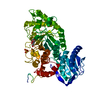
- PDB-3oh8: Crystal structure of the nucleoside-diphosphate sugar epimerase f... -
+ Open data

- Basic information
| Entry | Database: PDB / ID: 3oh8 | ||||||
|---|---|---|---|---|---|---|---|
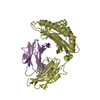
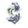
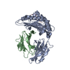
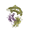
| Title | Crystal structure of the nucleoside-diphosphate sugar epimerase from Corynebacterium glutamicum. Northeast Structural Genomics Consortium Target CgR91 | ||||||
 Components Components | Nucleoside-diphosphate sugar epimerase (SulA family) | ||||||
 Keywords Keywords | ISOMERASE / DUF1731_C / Northeast Structural Genomics Consortium / NESG / CgR91 / PSI-Biology / Protein Structure Initiative | ||||||
| Function / homology | START domain / Alpha-D-Glucose-1,6-Bisphosphate; Chain A, domain 4 / NAD(P)-binding Rossmann-like Domain / Rossmann fold / 2-Layer Sandwich / 3-Layer(aba) Sandwich / Alpha Beta / :  Function and homology information Function and homology information | ||||||
| Biological species |  Corynebacterium glutamicum (bacteria) Corynebacterium glutamicum (bacteria) | ||||||
| Method |  X-RAY DIFFRACTION / SYNCHROTRON / SAD / Resolution: 1.997 Å X-RAY DIFFRACTION / SYNCHROTRON / SAD / Resolution: 1.997 Å | ||||||
 Authors Authors | Vorobiev, S. / Lew, S. / Kuzin, A. / Mao, M. / Xiao, R. / Ciccosanti, C. / Wang, H. / Everett, J.K. / Nair, R. / Acton, T.B. ...Vorobiev, S. / Lew, S. / Kuzin, A. / Mao, M. / Xiao, R. / Ciccosanti, C. / Wang, H. / Everett, J.K. / Nair, R. / Acton, T.B. / Rost, B. / Montelione, G.T. / Tong, L. / Hunt, J.F. / Northeast Structural Genomics Consortium (NESG) | ||||||
 Citation Citation | Journal: To be Published Title: Crystal structure of the nucleoside-diphosphate sugar epimerase from Corynebacterium glutamicum. Northeast Structural Genomics Consortium Target CgR91. Authors: Vorobiev, S. / Lew, S. / Kuzin, A. / Mao, M. / Xiao, R. / Ciccosanti, C. / Wang, H. / Everett, J.K. / Nair, R. / Acton, T.B. / Rost, B. / Montelione, G.T. / Tong, L. / Hunt, J.F. | ||||||
| History |
|
- Structure visualization
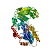
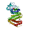
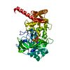
| Structure viewer | Molecule: MolmilJmol/JSmol |
|---|
- Downloads & links
-Download
| PDBx/mmCIF format | 3oh8.cif.gz | 187.6 KB | Display | PDBx/mmCIF format |
|---|---|---|---|---|
| PDB format | pdb3oh8.ent.gz | 149 KB | Display | PDB format |
| PDBx/mmJSON format | 3oh8.json.gz | Tree view | PDBx/mmJSON format | |
| Others |  Other downloads Other downloads |
-Validation report
| Arichive directory | https://data.pdbj.org/pub/pdb/validation_reports/oh/3oh8ftp://data.pdbj.org/pub/pdb/validation_reports/oh/3oh8 | HTTPS FTP |
|---|
-Related structure data
| Similar structure data | |
|---|---|
| Other databases |


-Links
 PDBj
PDBj- Assembly
| Deposited unit | 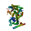
| ||||||||
|---|---|---|---|---|---|---|---|---|---|
| 1 |
| ||||||||
| Unit cell |
|
-Components
| #1: Protein | Mass: 56974.203 Da / Num. of mol.: 1 Source method: isolated from a genetically manipulated source Source: (gene. exp.) Corynebacterium glutamicum (bacteria) / Gene: cg1819 / Production host: |
|---|---|
| #2: Water | ChemComp-HOH /  Mass: 18.015 Da / Num. of mol.: 305 / Source method: isolated from a natural source / Formula: H2O Mass: 18.015 Da / Num. of mol.: 305 / Source method: isolated from a natural source / Formula: H2O |
| Has protein modification | Y |
-Experimental details
-Experiment
| Experiment | Method: X-RAY DIFFRACTION / Number of used crystals: 1 |
|---|
- Sample preparation
| Crystal | Density Matthews: 2.69 Å3/Da / Density % sol: 54.32 % |
|---|---|
| Crystal grow | Temperature: 278 K / Method: vapor diffusion, hanging drop / pH: 5 Details: 2% PEG 20000, 0.1M magnesium acetate, 0.1M sodium acetate, pH 5.0, VAPOR DIFFUSION, HANGING DROP, temperature 278K |
-Data collection
| Diffraction | Mean temperature: 100 K |
|---|---|
| Diffraction source | Source: SYNCHROTRON / Site: NSLS  / Beamline: X4A / Wavelength: 0.97901 Å / Beamline: X4A / Wavelength: 0.97901 Å |
| Detector | Type: ADSC QUANTUM 4r / Detector: CCD / Date: Aug 10, 2010 |
| Radiation | Monochromator: Si 111 CHANNEL / Protocol: SINGLE WAVELENGTH / Monochromatic (M) / Laue (L): M / Scattering type: x-ray |
| Radiation wavelength | Wavelength: 0.97901 Å / Relative weight: 1 |
| Reflection | Resolution: 2→50 Å / Num. all: 80580 / Num. obs: 73892 / % possible obs: 91.7 % / Observed criterion σ(F): 0 / Observed criterion σ(I): 0 / Redundancy: 7.2 % / Biso Wilson estimate: 23.1 Å2 / Rmerge(I) obs: 0.095 / Net I/σ(I): 22.03 |
| Reflection shell | Resolution: 2→2.07 Å / Redundancy: 7.1 % / Rmerge(I) obs: 0.508 / Mean I/σ(I) obs: 3.8 / Num. unique all: 8059 / % possible all: 99.9 |
- Processing
| Software |
| |||||||||||||||||||||||||||||||||||||||||||||||||||||||||||||||||||||||||||||||||||||||||||||||||||||||||||||||||||||||||||||||||||||||||||||||||||||||||||||||||||||||||||||||
|---|---|---|---|---|---|---|---|---|---|---|---|---|---|---|---|---|---|---|---|---|---|---|---|---|---|---|---|---|---|---|---|---|---|---|---|---|---|---|---|---|---|---|---|---|---|---|---|---|---|---|---|---|---|---|---|---|---|---|---|---|---|---|---|---|---|---|---|---|---|---|---|---|---|---|---|---|---|---|---|---|---|---|---|---|---|---|---|---|---|---|---|---|---|---|---|---|---|---|---|---|---|---|---|---|---|---|---|---|---|---|---|---|---|---|---|---|---|---|---|---|---|---|---|---|---|---|---|---|---|---|---|---|---|---|---|---|---|---|---|---|---|---|---|---|---|---|---|---|---|---|---|---|---|---|---|---|---|---|---|---|---|---|---|---|---|---|---|---|---|---|---|---|---|---|---|---|
| Refinement | Method to determine structure: SAD / Resolution: 1.997→38.776 Å / Occupancy max: 1 / Occupancy min: 1 / SU ML: 0.26 / Cross valid method: THROUGHOUT / σ(F): 1.03 / Phase error: 21.56 / Stereochemistry target values: ML
| |||||||||||||||||||||||||||||||||||||||||||||||||||||||||||||||||||||||||||||||||||||||||||||||||||||||||||||||||||||||||||||||||||||||||||||||||||||||||||||||||||||||||||||||
| Solvent computation | Shrinkage radii: 0.9 Å / VDW probe radii: 1.11 Å / Solvent model: FLAT BULK SOLVENT MODEL / Bsol: 40.556 Å2 / ksol: 0.331 e/Å3 | |||||||||||||||||||||||||||||||||||||||||||||||||||||||||||||||||||||||||||||||||||||||||||||||||||||||||||||||||||||||||||||||||||||||||||||||||||||||||||||||||||||||||||||||
| Displacement parameters | Biso max: 127.27 Å2 / Biso mean: 28.939 Å2 / Biso min: 7.75 Å2
| |||||||||||||||||||||||||||||||||||||||||||||||||||||||||||||||||||||||||||||||||||||||||||||||||||||||||||||||||||||||||||||||||||||||||||||||||||||||||||||||||||||||||||||||
| Refinement step | Cycle: LAST / Resolution: 1.997→38.776 Å
| |||||||||||||||||||||||||||||||||||||||||||||||||||||||||||||||||||||||||||||||||||||||||||||||||||||||||||||||||||||||||||||||||||||||||||||||||||||||||||||||||||||||||||||||
| Refine LS restraints |
| |||||||||||||||||||||||||||||||||||||||||||||||||||||||||||||||||||||||||||||||||||||||||||||||||||||||||||||||||||||||||||||||||||||||||||||||||||||||||||||||||||||||||||||||
| LS refinement shell | Refine-ID: X-RAY DIFFRACTION / Total num. of bins used: 24
| |||||||||||||||||||||||||||||||||||||||||||||||||||||||||||||||||||||||||||||||||||||||||||||||||||||||||||||||||||||||||||||||||||||||||||||||||||||||||||||||||||||||||||||||
| Refinement TLS params. | Method: refined / Origin x: 54.671 Å / Origin y: 27.233 Å / Origin z: 38.9902 Å
| |||||||||||||||||||||||||||||||||||||||||||||||||||||||||||||||||||||||||||||||||||||||||||||||||||||||||||||||||||||||||||||||||||||||||||||||||||||||||||||||||||||||||||||||
| Refinement TLS group | Selection details: chain A |
