 Movie
Movie Controller

[English] 日本語
 Yorodumi
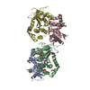
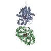
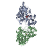
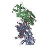
- PDB-6va0: Crystal structure of glucose-6-phosphate dehydrogenase W509A muta... -
+ Open data

- Basic information
| Entry | Database: PDB / ID: 6va0 | ||||||
|---|---|---|---|---|---|---|---|
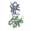
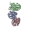
| Title | Crystal structure of glucose-6-phosphate dehydrogenase W509A mutant in complex with catalytic NADP+ | ||||||
 Components Components | Glucose-6-phosphate 1-dehydrogenase | ||||||
 Keywords Keywords | OXIDOREDUCTASE / Dehydrogenase / NADP+ | ||||||
| Function / homology |  Function and homology information Function and homology informationpentose biosynthetic process / ribose phosphate biosynthetic process / response to iron(III) ion / positive regulation of calcium ion transmembrane transport via high voltage-gated calcium channel / glucose-6-phosphate dehydrogenase (NADP+) / glucose-6-phosphate dehydrogenase activity / Pentose phosphate pathway / pentose-phosphate shunt, oxidative branch / negative regulation of cell growth involved in cardiac muscle cell development / glucose 6-phosphate metabolic process ...pentose biosynthetic process / ribose phosphate biosynthetic process / response to iron(III) ion / positive regulation of calcium ion transmembrane transport via high voltage-gated calcium channel / glucose-6-phosphate dehydrogenase (NADP+) / glucose-6-phosphate dehydrogenase activity / Pentose phosphate pathway / pentose-phosphate shunt, oxidative branch / negative regulation of cell growth involved in cardiac muscle cell development / glucose 6-phosphate metabolic process / NADP+ metabolic process / pentose-phosphate shunt / D-glucose binding / NFE2L2 regulates pentose phosphate pathway genes / Oxidoreductases / cholesterol biosynthetic process / erythrocyte maturation / response to food / negative regulation of reactive oxygen species metabolic process / substantia nigra development / regulation of neuron apoptotic process / TP53 Regulates Metabolic Genes / lipid metabolic process / glutathione metabolic process / glucose metabolic process / cytoplasmic side of plasma membrane / centriolar satellite / NADP binding / cellular response to oxidative stress / response to ethanol / protein homodimerization activity / extracellular exosome / membrane / identical protein binding / cytoplasm / cytosol Similarity search - Function | ||||||
| Biological species |  Homo sapiens (human) Homo sapiens (human) | ||||||
| Method |  X-RAY DIFFRACTION / SYNCHROTRON / MOLECULAR REPLACEMENT / molecular replacement / Resolution: 3.1 Å X-RAY DIFFRACTION / SYNCHROTRON / MOLECULAR REPLACEMENT / molecular replacement / Resolution: 3.1 Å | ||||||
 Authors Authors | Horikoshi, N. / Mochly-Rosen, D. / Wakatsuki, S. | ||||||
| Funding support |  United States, 1items United States, 1items
| ||||||
 Citation Citation | Journal: Proc.Natl.Acad.Sci.USA / Year: 2021 Title: Long-range structural defects by pathogenic mutations in most severe glucose-6-phosphate dehydrogenase deficiency. Authors: Horikoshi, N. / Hwang, S. / Gati, C. / Matsui, T. / Castillo-Orellana, C. / Raub, A.G. / Garcia, A.A. / Jabbarpour, F. / Batyuk, A. / Broweleit, J. / Xiang, X. / Chiang, A. / Broweleit, R. / ...Authors: Horikoshi, N. / Hwang, S. / Gati, C. / Matsui, T. / Castillo-Orellana, C. / Raub, A.G. / Garcia, A.A. / Jabbarpour, F. / Batyuk, A. / Broweleit, J. / Xiang, X. / Chiang, A. / Broweleit, R. / Vohringer-Martinez, E. / Mochly-Rosen, D. / Wakatsuki, S. | ||||||
| History |
|
- Structure visualization
| Structure viewer | Molecule: MolmilJmol/JSmol |
|---|
- Downloads & links
-Download
| PDBx/mmCIF format | 6va0.cif.gz | 229.4 KB | Display | PDBx/mmCIF format |
|---|---|---|---|---|
| PDB format | pdb6va0.ent.gz | 155.6 KB | Display | PDB format |
| PDBx/mmJSON format | 6va0.json.gz | Tree view | PDBx/mmJSON format | |
| Others |  Other downloads Other downloads |
-Validation report
| Arichive directory | https://data.pdbj.org/pub/pdb/validation_reports/va/6va0ftp://data.pdbj.org/pub/pdb/validation_reports/va/6va0 | HTTPS FTP |
|---|
-Related structure data
| Related structure data | 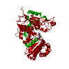 6va7C 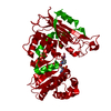 6va8C 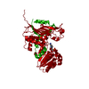 6va9C 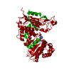 6vaqC  6a08S S: Starting model for refinement C: citing same article ( |
|---|---|
| Similar structure data |


-Links
 PDBj
PDBj- Assembly
| Deposited unit | 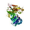
| ||||||||||
|---|---|---|---|---|---|---|---|---|---|---|---|
| 1 | 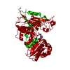
| ||||||||||
| Unit cell |
|
-Components
| #1: Protein | Mass: 59217.453 Da / Num. of mol.: 1 / Mutation: W509A Source method: isolated from a genetically manipulated source Source: (gene. exp.) Homo sapiens (human) / Gene: G6PD / Plasmid: pET28a / Production host:  References: UniProt: P11413, glucose-6-phosphate dehydrogenase (NADP+) |
|---|---|
| #2: Chemical | ChemComp-NAP /   Mass: 743.405 Da / Num. of mol.: 1 / Source method: obtained synthetically / Formula: C21H28N7O17P3 / Feature type: SUBJECT OF INVESTIGATION Mass: 743.405 Da / Num. of mol.: 1 / Source method: obtained synthetically / Formula: C21H28N7O17P3 / Feature type: SUBJECT OF INVESTIGATION |
| Has ligand of interest | Y |
-Experimental details
-Experiment
| Experiment | Method: X-RAY DIFFRACTION / Number of used crystals: 1 |
|---|
- Sample preparation
| Crystal | Density Matthews: 5.91 Å3/Da / Density % sol: 79.18 % |
|---|---|
| Crystal grow | Temperature: 293 K / Method: vapor diffusion, sitting drop / pH: 6.5 / Details: MES, PEG4000, Magnesium chloride, AG1 |
-Data collection
| Diffraction | Mean temperature: 100 K / Serial crystal experiment: N | ||||||||||||||||||||||||||||||||||||||||||||||||||||||||||||||||||||||||||||||||||||||||||||||||||||
|---|---|---|---|---|---|---|---|---|---|---|---|---|---|---|---|---|---|---|---|---|---|---|---|---|---|---|---|---|---|---|---|---|---|---|---|---|---|---|---|---|---|---|---|---|---|---|---|---|---|---|---|---|---|---|---|---|---|---|---|---|---|---|---|---|---|---|---|---|---|---|---|---|---|---|---|---|---|---|---|---|---|---|---|---|---|---|---|---|---|---|---|---|---|---|---|---|---|---|---|---|---|
| Diffraction source | Source: SYNCHROTRON / Site: ALS / Beamline: 5.0.2 / Wavelength: 0.99999 Å | ||||||||||||||||||||||||||||||||||||||||||||||||||||||||||||||||||||||||||||||||||||||||||||||||||||
| Detector | Type: DECTRIS PILATUS3 6M / Detector: PIXEL / Date: Aug 17, 2018 | ||||||||||||||||||||||||||||||||||||||||||||||||||||||||||||||||||||||||||||||||||||||||||||||||||||
| Radiation | Protocol: SINGLE WAVELENGTH / Monochromatic (M) / Laue (L): M / Scattering type: x-ray | ||||||||||||||||||||||||||||||||||||||||||||||||||||||||||||||||||||||||||||||||||||||||||||||||||||
| Radiation wavelength | Wavelength: 0.99999 Å / Relative weight: 1 | ||||||||||||||||||||||||||||||||||||||||||||||||||||||||||||||||||||||||||||||||||||||||||||||||||||
| Reflection | Resolution: 3.1→47.62 Å / Num. obs: 25692 / % possible obs: 97.2 % / Redundancy: 42.341 % / Biso Wilson estimate: 93.78 Å2 / CC1/2: 1 / Rmerge(I) obs: 0.143 / Rrim(I) all: 0.145 / Χ2: 1.072 / Net I/σ(I): 34.75 / Num. measured all: 1087829 | ||||||||||||||||||||||||||||||||||||||||||||||||||||||||||||||||||||||||||||||||||||||||||||||||||||
| Reflection shell | Diffraction-ID: 1
|
-Phasing
| Phasing | Method: molecular replacement |
|---|
- Processing
| Software |
| ||||||||||||||||||||||||||||||||||||||||||||||||||||||||||||||||||||||
|---|---|---|---|---|---|---|---|---|---|---|---|---|---|---|---|---|---|---|---|---|---|---|---|---|---|---|---|---|---|---|---|---|---|---|---|---|---|---|---|---|---|---|---|---|---|---|---|---|---|---|---|---|---|---|---|---|---|---|---|---|---|---|---|---|---|---|---|---|---|---|---|
| Refinement | Method to determine structure: MOLECULAR REPLACEMENT Starting model: 6A08 Resolution: 3.1→47.62 Å / SU ML: 0.3364 / Cross valid method: FREE R-VALUE / σ(F): 1.34 / Phase error: 26.0013 Stereochemistry target values: GeoStd + Monomer Library + CDL v1.2
| ||||||||||||||||||||||||||||||||||||||||||||||||||||||||||||||||||||||
| Solvent computation | Shrinkage radii: 0.9 Å / VDW probe radii: 1.11 Å / Solvent model: FLAT BULK SOLVENT MODEL | ||||||||||||||||||||||||||||||||||||||||||||||||||||||||||||||||||||||
| Displacement parameters | Biso mean: 94.46 Å2 | ||||||||||||||||||||||||||||||||||||||||||||||||||||||||||||||||||||||
| Refinement step | Cycle: LAST / Resolution: 3.1→47.62 Å
| ||||||||||||||||||||||||||||||||||||||||||||||||||||||||||||||||||||||
| Refine LS restraints |
| ||||||||||||||||||||||||||||||||||||||||||||||||||||||||||||||||||||||
| LS refinement shell |
| ||||||||||||||||||||||||||||||||||||||||||||||||||||||||||||||||||||||
| Refinement TLS params. | Method: refined / Origin x: 24.2464109505 Å / Origin y: 95.359487288 Å / Origin z: 30.1231071187 Å
| ||||||||||||||||||||||||||||||||||||||||||||||||||||||||||||||||||||||
| Refinement TLS group | Selection details: all |
