 Movie
Movie Controller

[English] 日本語
 Yorodumi
- PDB-3su6: Crystal structure of NS3/4A protease variant A156T in complex wit... -
+ Open data

- Basic information
| Entry | Database: PDB / ID: 3su6 | ||||||
|---|---|---|---|---|---|---|---|
| Title | Crystal structure of NS3/4A protease variant A156T in complex with vaniprevir | ||||||
 Components Components | NS3 protease, NS4A protein | ||||||
 Keywords Keywords | HYDROLASE/INHIBITOR / drug resistance / drug design / Protease inhibitors / serine protease / VIRAL PROTEIN / HYDROLASE-INHIBITOR complex | ||||||
| Function / homology |  Function and homology information Function and homology informationhost cell mitochondrial membrane / host cell lipid droplet / symbiont-mediated transformation of host cell / symbiont-mediated suppression of host TRAF-mediated signal transduction / symbiont-mediated perturbation of host cell cycle G1/S transition checkpoint / symbiont-mediated suppression of host JAK-STAT cascade via inhibition of STAT1 activity / symbiont-mediated suppression of host cytoplasmic pattern recognition receptor signaling pathway via inhibition of MAVS activity / SH3 domain binding / ribonucleoside triphosphate phosphatase activity / viral nucleocapsid ...host cell mitochondrial membrane / host cell lipid droplet / symbiont-mediated transformation of host cell / symbiont-mediated suppression of host TRAF-mediated signal transduction / symbiont-mediated perturbation of host cell cycle G1/S transition checkpoint / symbiont-mediated suppression of host JAK-STAT cascade via inhibition of STAT1 activity / symbiont-mediated suppression of host cytoplasmic pattern recognition receptor signaling pathway via inhibition of MAVS activity / SH3 domain binding / ribonucleoside triphosphate phosphatase activity / viral nucleocapsid / channel activity / monoatomic ion transmembrane transport / clathrin-dependent endocytosis of virus by host cell / RNA helicase activity / host cell perinuclear region of cytoplasm / host cell endoplasmic reticulum membrane / symbiont-mediated suppression of host type I interferon-mediated signaling pathway / ribonucleoprotein complex / serine-type endopeptidase activity / symbiont-mediated activation of host autophagy / cysteine-type endopeptidase activity / viral RNA genome replication / RNA-directed RNA polymerase activity / fusion of virus membrane with host endosome membrane / viral envelope / virion attachment to host cell / host cell plasma membrane / host cell nucleus / virion membrane / structural molecule activity / proteolysis / RNA binding / zinc ion binding / ATP binding Similarity search - Function | ||||||
| Biological species |  Hepatitis C virus Hepatitis C virus | ||||||
| Method |  X-RAY DIFFRACTION / SYNCHROTRON / MOLECULAR REPLACEMENT / Resolution: 1.1 Å X-RAY DIFFRACTION / SYNCHROTRON / MOLECULAR REPLACEMENT / Resolution: 1.1 Å | ||||||
 Authors Authors | Schiffer, C.A. / Romano, K.P. | ||||||
 Citation Citation | Journal: Plos Pathog. / Year: 2012 Title: The Molecular Basis of Drug Resistance against Hepatitis C Virus NS3/4A Protease Inhibitors. Authors: Romano, K.P. / Ali, A. / Aydin, C. / Soumana, D. / Ozen, A. / Deveau, L.M. / Silver, C. / Cao, H. / Newton, A. / Petropoulos, C.J. / Huang, W. / Schiffer, C.A. | ||||||
| History |
|
- Structure visualization
| Structure viewer | Molecule: MolmilJmol/JSmol |
|---|
- Downloads & links
-Download
| PDBx/mmCIF format | 3su6.cif.gz | 100.7 KB | Display | PDBx/mmCIF format |
|---|---|---|---|---|
| PDB format | pdb3su6.ent.gz | 74.6 KB | Display | PDB format |
| PDBx/mmJSON format | 3su6.json.gz | Tree view | PDBx/mmJSON format | |
| Others |  Other downloads Other downloads |
-Validation report
| Arichive directory | https://data.pdbj.org/pub/pdb/validation_reports/su/3su6ftp://data.pdbj.org/pub/pdb/validation_reports/su/3su6 | HTTPS FTP |
|---|
-Related structure data
| Related structure data |  3su0C  3su1C  3su2C  3su3C  3su4C  3su5C  3sudC  3sueC  3sufC  3sugC  3sv6C  3sv7C  3sv8C  3sv9C C: citing same article ( |
|---|---|
| Similar structure data |


-Links
 PDBj
PDBj
- Assembly
| Deposited unit | 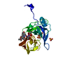
| ||||||||
|---|---|---|---|---|---|---|---|---|---|
| 1 |
| ||||||||
| Unit cell |
|
-Components
-Protein , 1 types, 1 molecules A
| #1: Protein | Mass: 21517.355 Da / Num. of mol.: 1 Fragment: NS4A (UNP residues 1674-1688), NS3 (UNP residues 1027-1208) Mutation: A1027S, P1028G, I1029D, L1039E, L1040E, I1043Q, I1044E, L1047Q, A1066T, C1073S, C1078L, I1098T, P1112Q, S1165A, C1185S, C1679S, V1686I, I1687N Source method: isolated from a genetically manipulated source Source: (gene. exp.) Hepatitis C virus / Strain: subtype 1a, BID-V318 / Gene: NS3-NS4A / Plasmid: PET28a / Production host:  |
|---|
-Non-polymers , 5 types, 199 molecules 


| #2: Chemical | ChemComp-SU3 / ( Mass: 757.936 Da / Num. of mol.: 1 / Source method: obtained synthetically / Formula: C38H55N5O9S Mass: 757.936 Da / Num. of mol.: 1 / Source method: obtained synthetically / Formula: C38H55N5O9S | ||||
|---|---|---|---|---|---|
| #3: Chemical | ChemComp-GOL /  Mass: 92.094 Da / Num. of mol.: 1 / Source method: obtained synthetically / Formula: C3H8O3 Mass: 92.094 Da / Num. of mol.: 1 / Source method: obtained synthetically / Formula: C3H8O3 | ||||
| #4: Chemical |  Mass: 96.063 Da / Num. of mol.: 2 / Source method: obtained synthetically / Formula: SO4 Mass: 96.063 Da / Num. of mol.: 2 / Source method: obtained synthetically / Formula: SO4#5: Chemical | ChemComp-ZN / |  Mass: 65.409 Da / Num. of mol.: 1 / Source method: obtained synthetically / Formula: Zn Mass: 65.409 Da / Num. of mol.: 1 / Source method: obtained synthetically / Formula: Zn#6: Water | ChemComp-HOH / | Mass: 18.015 Da / Num. of mol.: 194 / Source method: isolated from a natural source / Formula: H2O |
-Details
| Nonpolymer details | VANIPREVIR (MK-7009) IS A MACROCYCLIC, PEPTIDOMIMETIC HCV NS3/4A PROTEASE INHIBITOR FROM MERCK. THE ...VANIPREVIR |
|---|---|
| Sequence details | THE COFACTOR 4A RESIDUES 990-1000 (GLY SER VAL VAL ILE VAL GLY ARG ILE ASN LEU) IN THIS ENTRY ...THE COFACTOR 4A RESIDUES 990-1000 (GLY SER VAL VAL ILE VAL GLY ARG ILE ASN LEU) IN THIS ENTRY CORRESPOND |
-Experimental details
-Experiment
| Experiment | Method: X-RAY DIFFRACTION / Number of used crystals: 1 |
|---|
- Sample preparation
| Crystal | Density Matthews: 2.23 Å3/Da / Density % sol: 44.94 % |
|---|---|
| Crystal grow | Temperature: 295 K / Method: hanging drop, vapor diffusion / pH: 6.2 Details: 20-25% PEG 3350, 0.1M MES (pH 6.5), 4% ammonium sulfate, hanging drop, vapor diffusion, temperature 295K |
-Data collection
| Diffraction | Mean temperature: 100 K | |||||||||||||||||||||||||||||||||||||||||||||||||||||||||||||||||||||||||||||
|---|---|---|---|---|---|---|---|---|---|---|---|---|---|---|---|---|---|---|---|---|---|---|---|---|---|---|---|---|---|---|---|---|---|---|---|---|---|---|---|---|---|---|---|---|---|---|---|---|---|---|---|---|---|---|---|---|---|---|---|---|---|---|---|---|---|---|---|---|---|---|---|---|---|---|---|---|---|---|
| Diffraction source | Source: SYNCHROTRON / Site: APS  / Beamline: 21-ID-F / Wavelength: 0.97872 Å / Beamline: 21-ID-F / Wavelength: 0.97872 Å | |||||||||||||||||||||||||||||||||||||||||||||||||||||||||||||||||||||||||||||
| Detector | Date: Dec 7, 2010 | |||||||||||||||||||||||||||||||||||||||||||||||||||||||||||||||||||||||||||||
| Radiation | Protocol: SINGLE WAVELENGTH / Scattering type: x-ray | |||||||||||||||||||||||||||||||||||||||||||||||||||||||||||||||||||||||||||||
| Radiation wavelength | Wavelength: 0.97872 Å / Relative weight: 1 | |||||||||||||||||||||||||||||||||||||||||||||||||||||||||||||||||||||||||||||
| Reflection | Resolution: 1.1→50 Å / Num. obs: 76300 / % possible obs: 96.6 % / Redundancy: 5.2 % / Rmerge(I) obs: 0.058 / Χ2: 1.001 / Net I/σ(I): 12.1 | |||||||||||||||||||||||||||||||||||||||||||||||||||||||||||||||||||||||||||||
| Reflection shell |
|
- Processing
| Software |
| ||||||||||||||||||||||||||||||||||||||||||||||||||||||||||||||||||||||||||||||||||||||||||
|---|---|---|---|---|---|---|---|---|---|---|---|---|---|---|---|---|---|---|---|---|---|---|---|---|---|---|---|---|---|---|---|---|---|---|---|---|---|---|---|---|---|---|---|---|---|---|---|---|---|---|---|---|---|---|---|---|---|---|---|---|---|---|---|---|---|---|---|---|---|---|---|---|---|---|---|---|---|---|---|---|---|---|---|---|---|---|---|---|---|---|---|
| Refinement | Method to determine structure: MOLECULAR REPLACEMENT / Resolution: 1.1→33.28 Å / Cor.coef. Fo:Fc: 0.971 / Cor.coef. Fo:Fc free: 0.969 / WRfactor Rfree: 0.1762 / WRfactor Rwork: 0.1569 / Occupancy max: 1 / Occupancy min: 0.5 / FOM work R set: 0.9201 / SU B: 0.756 / SU ML: 0.017 / SU R Cruickshank DPI: 0.0297 / SU Rfree: 0.029 / Cross valid method: THROUGHOUT / σ(F): 0 / ESU R Free: 0.029 / Stereochemistry target values: MAXIMUM LIKELIHOOD Details: HYDROGENS HAVE BEEN ADDED IN THE RIDING POSITIONS U VALUES: REFINED INDIVIDUALLY
| ||||||||||||||||||||||||||||||||||||||||||||||||||||||||||||||||||||||||||||||||||||||||||
| Solvent computation | Ion probe radii: 0.8 Å / Shrinkage radii: 0.8 Å / VDW probe radii: 1.4 Å / Solvent model: BABINET MODEL WITH MASK | ||||||||||||||||||||||||||||||||||||||||||||||||||||||||||||||||||||||||||||||||||||||||||
| Displacement parameters | Biso max: 96.58 Å2 / Biso mean: 12.9318 Å2 / Biso min: 4.9 Å2
| ||||||||||||||||||||||||||||||||||||||||||||||||||||||||||||||||||||||||||||||||||||||||||
| Refinement step | Cycle: LAST / Resolution: 1.1→33.28 Å
| ||||||||||||||||||||||||||||||||||||||||||||||||||||||||||||||||||||||||||||||||||||||||||
| Refine LS restraints |
| ||||||||||||||||||||||||||||||||||||||||||||||||||||||||||||||||||||||||||||||||||||||||||
| LS refinement shell | Resolution: 1.1→1.128 Å / Total num. of bins used: 20
|
