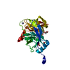
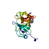
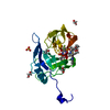
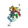
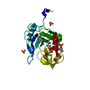
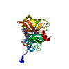
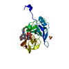
[English] 日本語
Yorodumi
- PDB-6c2n: Crystal structure of HCV NS3/4A double mutant variant Y56H/D168A ... -

+
Open data


ID or keywords:

Loading...

-
Basic information

Entry
Database: PDB / ID: 6c2n
TitleCrystal structure of HCV NS3/4A double mutant variant Y56H/D168A in complex with danoprevir
ComponentsNS4A COFACTOR-NS3 PROTEIN CHIMERA
KeywordsHYDROLASE / NS3/4a Protease / Hepatitis C virus / Drug Resistance / protease inhibitor / HYDROLASE-HYDROLASE INHIBITOR complex
Function / homology
Function and homology information


host cell mitochondrial membrane / host cell lipid droplet / symbiont-mediated suppression of host TRAF-mediated signal transduction / transformation of host cell by virus / symbiont-mediated perturbation of host cell cycle G1/S transition checkpoint / symbiont-mediated suppression of host JAK-STAT cascade via inhibition of STAT1 activity / symbiont-mediated suppression of host cytoplasmic pattern recognition receptor signaling pathway via inhibition of MAVS activity / ribonucleoside triphosphate phosphatase activity / lipid droplet / protein complex oligomerization ...host cell mitochondrial membrane / host cell lipid droplet / symbiont-mediated suppression of host TRAF-mediated signal transduction / transformation of host cell by virus / symbiont-mediated perturbation of host cell cycle G1/S transition checkpoint / symbiont-mediated suppression of host JAK-STAT cascade via inhibition of STAT1 activity / symbiont-mediated suppression of host cytoplasmic pattern recognition receptor signaling pathway via inhibition of MAVS activity / ribonucleoside triphosphate phosphatase activity / lipid droplet / protein complex oligomerization / monoatomic ion channel activity / viral nucleocapsid / clathrin-dependent endocytosis of virus by host cell / RNA helicase activity / host cell endoplasmic reticulum membrane / host cell perinuclear region of cytoplasm / symbiont-mediated suppression of host type I interferon-mediated signaling pathway / ribonucleoprotein complex / induction by virus of host autophagy / viral RNA genome replication / cysteine-type endopeptidase activity / serine-type endopeptidase activity / RNA-dependent RNA polymerase activity / virus-mediated perturbation of host defense response / fusion of virus membrane with host endosome membrane / viral envelope / virion attachment to host cell / host cell plasma membrane / structural molecule activity / virion membrane / proteolysis / RNA binding / zinc ion binding / ATP binding / plasma membrane / cytoplasm
Similarity search - Function
Thrombin, subunit H - #120 / : / Hepatitis C virus core protein, chain A superfamily / Hepatitus C virus, Non-structural 5a protein, C-terminal / Hepatitis C virus NS5A, 1B domain superfamily / Hepatitis C virus non-structural protein NS2, C-terminal domain / Hepatitis C virus non-structural protein NS2, N-terminal domain / Hepatitis C virus non-structural protein NS2 / HCV NS5a protein C-terminal region / Hepatitis C virus, Non-structural protein NS4b ...Thrombin, subunit H - #120 / : / Hepatitis C virus core protein, chain A superfamily / Hepatitus C virus, Non-structural 5a protein, C-terminal / Hepatitis C virus NS5A, 1B domain superfamily / Hepatitis C virus non-structural protein NS2, C-terminal domain / Hepatitis C virus non-structural protein NS2, N-terminal domain / Hepatitis C virus non-structural protein NS2 / HCV NS5a protein C-terminal region / Hepatitis C virus, Non-structural protein NS4b / Hepatitis C virus, Core protein, N-terminal / Hepatitis C virus non-structural protein NS4b / Hepatitis C virus capsid protein / Hepatitis C virus, Non-structural protein NS2 / Hepatitis C virus, Non-structural 5a protein / Hepatitis C virus, Non-structural 5a protein, domain 1a / Hepatitis C virus non-structural 5a, 1B domain / NS5A domain 1a superfamily / Hepatitis C virus non-structural 5a protein membrane anchor / Hepatitis C virus non-structural 5a zinc finger domain / Hepatitis C virus non-structural 5a domain 1b / Hepacivirus nonstructural protein 2 (NS2) protease domain profile. / Hepatitis C virus, Non-structural protein NS4a / Hepatitis C virus non-structural protein NS4a / Hepatitis C virus, Core protein, C-terminal / Hepatitis C virus core protein / Hepatitis C virus, Non-structural protein E2/NS1 / Hepatitis C virus non-structural protein E2/NS1 / Hepatitis C virus, Envelope glycoprotein E1 / Hepatitis C virus envelope glycoprotein E1 / RNA dependent RNA polymerase, hepatitis C virus / Viral RNA dependent RNA polymerase / Hepatitis C virus, NS3 protease, Peptidase S29 / Hepatitis C virus NS3 protease / Hepacivirus/Pegivirus NS3 protease domain profile. / DEAD box, Flavivirus / Flavivirus DEAD domain / helicase superfamily c-terminal domain / Trypsin-like serine proteases / Superfamilies 1 and 2 helicase C-terminal domain profile. / Superfamilies 1 and 2 helicase ATP-binding type-1 domain profile. / DEAD-like helicases superfamily / Helicase, C-terminal / Helicase superfamily 1/2, ATP-binding domain / Thrombin, subunit H / Reverse transcriptase/Diguanylate cyclase domain / RNA-directed RNA polymerase, catalytic domain / RdRp of positive ssRNA viruses catalytic domain profile. / Peptidase S1, PA clan, chymotrypsin-like fold / Peptidase S1, PA clan / DNA/RNA polymerase superfamily / Beta Barrel / P-loop containing nucleoside triphosphate hydrolase / Mainly Beta
Similarity search - Domain/homology
Chem-TSV / Genome polyprotein
Similarity search - Component
Biological speciesHepatitis C virus subtype 1a
MethodX-RAY DIFFRACTION / MOLECULAR REPLACEMENT / Resolution: 1.802 Å
AuthorsMatthew, A.N. / Schiffer, C.A.
Funding support United States, 2items
OrganizationGrant numberCountry
National Institutes of Health/National Institute Of Allergy and Infectious Diseases (NIH/NIAID)R01 AI085051 United States
National Institutes of Health/National Institute of General Medical Sciences (NIH/NIGMS)F31 GM119345 United States
CitationJournal: To Be Published
Title: Clinical signature variant of HCV NS3/4A protease uses a novel mechanism to confer resistance
Authors: Matthew, A.N. / Schiffer, C.A.
History
DepositionJan 8, 2018Deposition site: RCSB / Processing site: RCSB
Revision 1.0Jan 16, 2019Provider: repository / Type: Initial release
Revision 1.1Feb 20, 2019Group: Author supporting evidence / Data collection / Category: pdbx_audit_support / Item: _pdbx_audit_support.funding_organization
Revision 1.2Dec 18, 2019Group: Author supporting evidence / Category: pdbx_audit_support / Item: _pdbx_audit_support.funding_organization
Revision 1.3Oct 4, 2023Group: Data collection / Database references ...Data collection / Database references / Derived calculations / Refinement description / Structure summary
Category: chem_comp / chem_comp_atom ...chem_comp / chem_comp_atom / chem_comp_bond / database_2 / entity / pdbx_entity_nonpoly / pdbx_initial_refinement_model
Item: _chem_comp.name / _database_2.pdbx_DOI ..._chem_comp.name / _database_2.pdbx_DOI / _database_2.pdbx_database_accession / _entity.pdbx_description / _pdbx_entity_nonpoly.name

-
Structure visualization

Structure viewerMolecule:
MolmilJmol/JSmol

Downloads & links

-
Assembly

Deposited unit
A: NS4A COFACTOR-NS3 PROTEIN CHIMERA
hetero molecules


Theoretical massNumber of molelcules
Total (without water)22,3264
Polymers21,4341
Non-polymers8913
Water3,567198
1


  • Idetical with deposited unit
  • defined by author&software
  • Evidence: gel filtration
TypeNameSymmetry operationNumber
identity operation1_555x,y,z1
MethodPISA
Unit cell
Length a, b, c (Å)59.964, 55.355, 58.891
Angle α, β, γ (deg.)90.00, 90.00, 90.00
Int Tables number19
Space group name H-MP212121

-
Components

#1: Protein NS4A COFACTOR-NS3 PROTEIN CHIMERA


Mass: 21434.293 Da / Num. of mol.: 1
Source method: isolated from a genetically manipulated source
Source: (gene. exp.) Hepatitis C virus subtype 1a / Plasmid: pET-28a / Production host: Escherichia coli BL21(DE3) (bacteria) / Strain (production host): BL21(DE3) / References: UniProt: A8DG50
#2: Chemical ChemComp-ZN / ZINC ION


Mass: 65.409 Da / Num. of mol.: 1 / Source method: obtained synthetically / Formula: Zn
#3: Chemical ChemComp-SO4 / SULFATE ION


Mass: 96.063 Da / Num. of mol.: 1 / Source method: obtained synthetically / Formula: SO4
#4: Chemical ChemComp-TSV / (2R,6S,12Z,13aS,14aR,16aS)-6-[(tert-butoxycarbonyl)amino]-14a-[(cyclopropylsulfonyl)carbamoyl]-5,16-dioxo-1,2,3,5,6,7,8 ,9,10,11,13a,14,14a,15,16,16a-hexadecahydrocyclopropa[e]pyrrolo[1,2-a][1,4]diazacyclopentadecin-2-yl 4-fluoro-2H-isoindole-2-carboxylate / ITMN-191 / danoprevir


Mass: 729.815 Da / Num. of mol.: 1 / Source method: obtained synthetically / Formula: C35H44FN5O9S / Comment: inhibitor*YM
#5: Water ChemComp-HOH / water


Mass: 18.015 Da / Num. of mol.: 198 / Source method: isolated from a natural source / Formula: H2O

-
Experimental details

-
Experiment

ExperimentMethod: X-RAY DIFFRACTION / Number of used crystals: 1

-
Sample preparation

CrystalDensity Matthews: 2.3 Å3/Da / Density % sol: 46.41 %
Crystal growTemperature: 288 K / Method: vapor diffusion, hanging drop
Details: 100 mM MES BUFFER PH 6.5, 4% (W/V) AMMONIUM SULFATE, 20-26% PEG 3350

-
Data collection

DiffractionMean temperature: 100 K
Diffraction sourceSource: ROTATING ANODE / Type: RIGAKU MICROMAX-007 HF / Wavelength: 1.54178 Å
DetectorType: RIGAKU SATURN 944 / Detector: CCD / Date: Jul 25, 2017
RadiationProtocol: SINGLE WAVELENGTH / Monochromatic (M) / Laue (L): M / Scattering type: x-ray
Radiation wavelengthWavelength: 1.54178 Å / Relative weight: 1
ReflectionResolution: 1.802→27.677 Å / Num. obs: 18030 / % possible obs: 96.4 % / Redundancy: 7.7 % / Rsym value: 0.098 / Net I/σ(I): 17.8
Reflection shellResolution: 1.802→1.914 Å / Rsym value: 0.299

-
Processing

Software
NameVersionClassification
HKL-3000703xdata scaling
PHASERphasing
PHENIX1.12-2829refinement
Coot0.8.8model building
HKL-3000data collection
HKL-3000data processing
RefinementMethod to determine structure: MOLECULAR REPLACEMENT
Starting model: 5voj
Resolution: 1.802→27.677 Å / SU ML: 0.17 / Cross valid method: FREE R-VALUE / σ(F): 1.34 / Phase error: 17.97
RfactorNum. reflection% reflection
Rfree0.1897 866 4.82 %
Rwork0.1624 --
obs0.1638 17982 96.06 %
Solvent computationShrinkage radii: 0.9 Å / VDW probe radii: 1.11 Å
Refinement stepCycle: LAST / Resolution: 1.802→27.677 Å
ProteinNucleic acidLigandSolventTotal
Num. atoms1421 0 57 198 1676
Refine LS restraints
Refine-IDTypeDev idealNumber
X-RAY DIFFRACTIONf_bond_d0.0091514
X-RAY DIFFRACTIONf_angle_d1.3032074
X-RAY DIFFRACTIONf_dihedral_angle_d16.918894
X-RAY DIFFRACTIONf_chiral_restr0.068244
X-RAY DIFFRACTIONf_plane_restr0.008263
LS refinement shell
Resolution (Å)Rfactor RfreeNum. reflection RfreeRfactor RworkNum. reflection RworkRefine-ID% reflection obs (%)
1.8015-1.91440.23591150.19992242X-RAY DIFFRACTION77
1.9144-2.06210.1981480.16022910X-RAY DIFFRACTION100
2.0621-2.26960.18031480.14832931X-RAY DIFFRACTION100
2.2696-2.59780.18921470.15152946X-RAY DIFFRACTION100
2.5978-3.27210.18011480.16562983X-RAY DIFFRACTION100
3.2721-27.68080.18891600.1653104X-RAY DIFFRACTION100

+
About Yorodumi

-
News

-
Feb 9, 2022. New format data for meta-information of EMDB entries

New format data for meta-information of EMDB entries

  • Version 3 of the EMDB header file is now the official format.
  • The previous official version 1.9 will be removed from the archive.

Related info.:EMDB header

External links:wwPDB to switch to version 3 of the EMDB data model

-
Aug 12, 2020. Covid-19 info

Covid-19 info

URL: https://pdbjlvh1.pdbj.org/emnavi/covid19.php

New page: Covid-19 featured information page in EM Navigator.

Related info.:Covid-19 info / Mar 5, 2020. Novel coronavirus structure data

+
Mar 5, 2020. Novel coronavirus structure data

Novel coronavirus structure data

Related info.:Yorodumi Speices / Aug 12, 2020. Covid-19 info

External links:COVID-19 featured content - PDBj / Molecule of the Month (242):Coronavirus Proteases

+
Jan 31, 2019. EMDB accession codes are about to change! (news from PDBe EMDB page)

EMDB accession codes are about to change! (news from PDBe EMDB page)

  • The allocation of 4 digits for EMDB accession codes will soon come to an end. Whilst these codes will remain in use, new EMDB accession codes will include an additional digit and will expand incrementally as the available range of codes is exhausted. The current 4-digit format prefixed with “EMD-” (i.e. EMD-XXXX) will advance to a 5-digit format (i.e. EMD-XXXXX), and so on. It is currently estimated that the 4-digit codes will be depleted around Spring 2019, at which point the 5-digit format will come into force.
  • The EM Navigator/Yorodumi systems omit the EMD- prefix.

Related info.:Q: What is EMD? / ID/Accession-code notation in Yorodumi/EM Navigator

External links:EMDB Accession Codes are Changing Soon! / Contact to PDBj

+
Jul 12, 2017. Major update of PDB

Major update of PDB

  • wwPDB released updated PDB data conforming to the new PDBx/mmCIF dictionary.
  • This is a major update changing the version number from 4 to 5, and with Remediation, in which all the entries are updated.
  • In this update, many items about electron microscopy experimental information are reorganized (e.g. em_software).
  • Now, EM Navigator and Yorodumi are based on the updated data.

External links:wwPDB Remediation / Enriched Model Files Conforming to OneDep Data Standards Now Available in the PDB FTP Archive

-
Yorodumi

Thousand views of thousand structures

  • Yorodumi is a browser for structure data from EMDB, PDB, SASBDB, etc.
  • This page is also the successor to EM Navigator detail page, and also detail information page/front-end page for Omokage search.
  • The word "yorodu" (or yorozu) is an old Japanese word meaning "ten thousand". "mi" (miru) is to see.

Related info.:EMDB / PDB / SASBDB / Comparison of 3 databanks / Yorodumi Search / Aug 31, 2016. New EM Navigator & Yorodumi / Yorodumi Papers / Jmol/JSmol / Function and homology information / Changes in new EM Navigator and Yorodumi

Read more