 Movie
Movie Controller

[English] 日本語
 Yorodumi
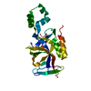
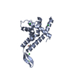
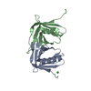
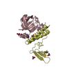
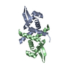
- PDB-3ssi: PROTEINASE INHIBITOR SSI (STREPTOMYCES SUBTILISIN, INHIBITOR) FRO... -
+ Open data

- Basic information
| Entry | Database: PDB / ID: 3ssi | |||||||||
|---|---|---|---|---|---|---|---|---|---|---|
| Title | PROTEINASE INHIBITOR SSI (STREPTOMYCES SUBTILISIN, INHIBITOR) FROM STREPTOMYCES ALBOGRISEOLUS | |||||||||
 Components Components | STREPTOMYCES SUBTILISIN INHIBITOR | |||||||||
 Keywords Keywords | SERINE PROTEASE INHIBITOR / SSI / SUBTILISIN BPN | |||||||||
| Function / homology |  Function and homology information Function and homology information | |||||||||
| Biological species |  Streptomyces albogriseolus (bacteria) Streptomyces albogriseolus (bacteria) | |||||||||
| Method |  X-RAY DIFFRACTION / Resolution: 2.3 Å X-RAY DIFFRACTION / Resolution: 2.3 Å | |||||||||
 Authors Authors | Suzuki, T. / Nonaka, T. / Mitsui, Y. | |||||||||
 Citation Citation | Journal: To be Published Title: Structural Modulation of the Protein Proteinase Inhibitor Ssi (Streptomyces Subtilisin Inhibitor) Authors: Suzuki, T. #1: Journal: Proc.Natl.Acad.Sci.USA / Year: 1992Title: Crystal Structure of an Engineered Subtilisin Inhibitor Complexed with Bovine Trypsin Authors: Takeuchi, Y. / Nonaka, T. / Nakamura, K.T. / Kojima, S. / Miura, K. / Mitsui, Y. #2: Journal: J.Mol.Biol. / Year: 1991Title: Refined Crystal Structure of the Complex of Subtilisin Bpn' and Streptomyces Subtilisin Inhibitor at 1.8 A Resolution Authors: Takeuchi, Y. / Satow, Y. / Nakamura, K.T. / Mitsui, Y. #3: Journal: Protein Eng. / Year: 1991Title: Molecular Recognition at the Active Site of Subtilisin Bpn': Crystallographic Studies Using Genetically Engineered Proteinaceous Inhibitor Ssi (Streptomyces Subtilisin Inhibitor) Authors: Takeuchi, Y. / Noguchi, S. / Satow, Y. / Kojima, S. / Kumagai, I. / Miura, K. / Nakamura, K.T. / Mitsui, Y. #4: Journal: J.Biochem.(Tokyo) / Year: 1985Title: Interactions of Streptomyces Subtilisin Inhibitor with Streptomyces Griseus Proteases a and B. Enzyme Kinetic and Computer Simulation Studies Authors: Christensen, U. / Ishida, S. / Ishii, S. / Mitsui, Y. / Iitaka, Y. / Mcclarin, J. / Langridge, R. #6: Journal: J.Mol.Biol. / Year: 1984Title: Crystal Structure at 2.6 A Resolution of the Complex of Subtilisin Bpn' with Streptomyces Subtilisin Inhibitor Authors: Hirono, S. / Akagawa, H. / Mitsui, Y. / Iitaka, Y. #7: Journal: J.Biochem.(Tokyo) / Year: 1980Title: Solvent Accessibility and Microenvironment in a Bacterial Protein Proteinase Inhibitor Ssi (Streptomyces Subtilisin Inhibitor) Authors: Satow, Y. / Watanabe, Y. / Mitsui, Y. #8: Journal: J.Mol.Biol. / Year: 1979Title: Crystal Structure of the Complex of Subtilisin Bpn' with its Protein Inhibitor Streptomyces Subtilisin Inhibitor. The Structure at 4.3 Angstroms Resolution Authors: Hirono, S. / Nakamura, K.T. / Iitaka, Y. / Mitsui, Y. #9: Journal: J.Mol.Biol. / Year: 1979Title: Crystal Structure of a Bacterial Protein Proteinase Inhibitor (Streptomyces Subtilisin Inhibitor) at 2.6 A Resolution Authors: Mitsui, Y. / Satow, Y. / Watanabe, Y. / Iitaka, Y. #10: Journal: Nature / Year: 1979Title: Crystal Structures of Streptomyces Subtilisin Inhibitor and its Complex with Subtilisin Bpn Authors: Mitsui, Y. / Satow, Y. / Watanabe, Y. / Hirono, S. / Iitaka, Y. #11: Journal: J.Biochem.(Tokyo) / Year: 1978Title: Crystal Structure of a Protein Proteinase Inhibitor, Ssi (Streptomyces Subtilisin Inhibitor), at 4 A Resolution Authors: Satow, Y. / Mitsui, Y. / Iitaka, Y. #12: Journal: J.Biochem.(Tokyo) / Year: 1977Title: Crystal Structure of a Protein Proteinase Inhibitor, Streptomyces Subtilisin Inhibitor, at 2.3 Angstrom Resolution Authors: Mitsui, Y. / Satow, Y. / Sakamaki, T. / Iitaka, Y. #13: Journal: J.Mol.Biol. / Year: 1973Title: Crystallization and Preliminary X-Ray Investigation of a New Alkaline Protease Inhibitor and its Complex with Subtilisin Bpn Authors: Satow, Y. / Mitsui, Y. / Iitaka, Y. / Murao, S. / Sato, S. | |||||||||
| History |
|
- Structure visualization
| Structure viewer | Molecule: MolmilJmol/JSmol |
|---|
- Downloads & links
-Download
| PDBx/mmCIF format | 3ssi.cif.gz | 32.7 KB | Display | PDBx/mmCIF format |
|---|---|---|---|---|
| PDB format | pdb3ssi.ent.gz | 21.5 KB | Display | PDB format |
| PDBx/mmJSON format | 3ssi.json.gz | Tree view | PDBx/mmJSON format | |
| Others |  Other downloads Other downloads |
-Validation report
| Arichive directory | https://data.pdbj.org/pub/pdb/validation_reports/ss/3ssiftp://data.pdbj.org/pub/pdb/validation_reports/ss/3ssi | HTTPS FTP |
|---|
-Related structure data
| Similar structure data |
|---|

-Links
 PDBj
PDBj- Assembly
| Deposited unit | 
| ||||||||
|---|---|---|---|---|---|---|---|---|---|
| 1 | 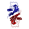
| ||||||||
| Unit cell |
|
-Components
| #1: Protein | Mass: 11494.929 Da / Num. of mol.: 1 / Source method: isolated from a natural source / Source: (natural) Streptomyces albogriseolus (bacteria) / Strain: S-3253 / References: UniProt: P01006 |
|---|---|
| #2: Water | ChemComp-HOH /  Mass: 18.015 Da / Num. of mol.: 31 / Source method: isolated from a natural source / Formula: H2O Mass: 18.015 Da / Num. of mol.: 31 / Source method: isolated from a natural source / Formula: H2O |
| Has protein modification | Y |
-Experimental details
-Experiment
| Experiment | Method: X-RAY DIFFRACTION / Number of used crystals: 1 |
|---|
- Sample preparation
| Crystal | Density Matthews: 2.4 Å3/Da / Density % sol: 49 % |
|---|---|
| Crystal grow | pH: 7 / Details: pH 7.0 |
-Data collection
| Diffraction | Mean temperature: 293 K |
|---|---|
| Diffraction source | Source: ROTATING ANODE / Type: RIGAKU RUH2R / Wavelength: 1.5418 |
| Detector | Type: RIGAKU / Detector: IMAGE PLATE / Date: Feb 26, 1993 / Details: SUPPER DOUBLE FOCUSING |
| Radiation | Monochromatic (M) / Laue (L): M / Scattering type: x-ray |
| Radiation wavelength | Wavelength: 1.5418 Å / Relative weight: 1 |
| Reflection | Resolution: 2.03→38.3 Å / Num. obs: 4866 / % possible obs: 63.7 % / Observed criterion σ(I): 1 / Redundancy: 2.6 % / Rmerge(I) obs: 0.048 |
| Reflection shell | Resolution: 2.3→2.34 Å / % possible all: 48.8 |
- Processing
| Software |
| ||||||||||||||||||||||||||||||||||||||||||||||||||||||||||||
|---|---|---|---|---|---|---|---|---|---|---|---|---|---|---|---|---|---|---|---|---|---|---|---|---|---|---|---|---|---|---|---|---|---|---|---|---|---|---|---|---|---|---|---|---|---|---|---|---|---|---|---|---|---|---|---|---|---|---|---|---|---|
| Refinement | Resolution: 2.3→6 Å / σ(F): 2
| ||||||||||||||||||||||||||||||||||||||||||||||||||||||||||||
| Displacement parameters | Biso mean: 31.8 Å2 | ||||||||||||||||||||||||||||||||||||||||||||||||||||||||||||
| Refine analyze | Luzzati coordinate error obs: 0.15 Å | ||||||||||||||||||||||||||||||||||||||||||||||||||||||||||||
| Refinement step | Cycle: LAST / Resolution: 2.3→6 Å
| ||||||||||||||||||||||||||||||||||||||||||||||||||||||||||||
| Refine LS restraints |
|
