 Movie
Movie Controller

[English] 日本語
 Yorodumi
- PDB-3snp: Crystal structure analysis of iron regulatory protein 1 in comple... -
+ Open data

- Basic information
| Entry | Database: PDB / ID: 3snp | |||||||||
|---|---|---|---|---|---|---|---|---|---|---|
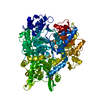
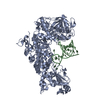
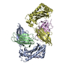
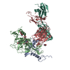
| Title | Crystal structure analysis of iron regulatory protein 1 in complex with ferritin H IRE RNA | |||||||||
 Components Components |
| |||||||||
 Keywords Keywords | LYASE/RNA / RNA binding / iron sulfur cluster binding / phosphorylation / LYASE-RNA complex | |||||||||
| Function / homology |  Function and homology information Function and homology informationaconitate hydratase / aconitate hydratase activity / iron-responsive element binding / citrate metabolic process / response to iron(II) ion / tricarboxylic acid cycle / 4 iron, 4 sulfur cluster binding / intracellular iron ion homeostasis / metal ion binding / cytosol Similarity search - Function | |||||||||
| Biological species |  | |||||||||
| Method |  X-RAY DIFFRACTION / SYNCHROTRON / MOLECULAR REPLACEMENT / Resolution: 2.8 Å X-RAY DIFFRACTION / SYNCHROTRON / MOLECULAR REPLACEMENT / Resolution: 2.8 Å | |||||||||
 Authors Authors | Volz, K. / Selezneva, A.I. / Walden, W.E. | |||||||||
 Citation Citation | Journal: Science / Year: 2006 Title: Structure of dual function iron regulatory protein 1 complexed with ferritin IRE-RNA. Authors: Walden, W.E. / Selezneva, A.I. / Dupuy, J. / Volbeda, A. / Fontecilla-Camps, J.C. / Theil, E.C. / Volz, K. #1: Journal: Acta Crystallogr.,Sect.F / Year: 2006 Title: Crystallization and preliminary X-ray diffraction analysis of iron regulatory protein 1 in complex with ferritin IRE RNA. Authors: Selezneva, A.I. / Cavigiolio, G. / Theil, E.C. / Walden, W.E. / Volz, K. | |||||||||
| History |
|
- Structure visualization
| Structure viewer | Molecule: MolmilJmol/JSmol |
|---|
- Downloads & links
-Download
| PDBx/mmCIF format | 3snp.cif.gz | 387.3 KB | Display | PDBx/mmCIF format |
|---|---|---|---|---|
| PDB format | pdb3snp.ent.gz | 304.9 KB | Display | PDB format |
| PDBx/mmJSON format | 3snp.json.gz | Tree view | PDBx/mmJSON format | |
| Others |  Other downloads Other downloads |
-Validation report
| Arichive directory | https://data.pdbj.org/pub/pdb/validation_reports/sn/3snpftp://data.pdbj.org/pub/pdb/validation_reports/sn/3snp | HTTPS FTP |
|---|
-Related structure data


-Links
 PDBj
PDBj


- Assembly
| Deposited unit | 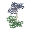
| ||||||||
|---|---|---|---|---|---|---|---|---|---|
| 1 | 
| ||||||||
| 2 | 
| ||||||||
| Unit cell |
|
-Components
| #1: Protein | Mass: 100776.734 Da / Num. of mol.: 2 / Mutation: C437S/C503S Source method: isolated from a genetically manipulated source Source: (gene. exp.)  #2: RNA chain | Mass: 9568.674 Da / Num. of mol.: 2 / Source method: obtained synthetically #3: Water | ChemComp-HOH / |  Mass: 18.015 Da / Num. of mol.: 557 / Source method: isolated from a natural source / Formula: H2O Mass: 18.015 Da / Num. of mol.: 557 / Source method: isolated from a natural source / Formula: H2OSequence details | THE AUTHORS STATE THAT THE SEQUENCE PROVIDED FOR CHAINS A AND B IS CORRECT. | |
|---|
-Experimental details
-Experiment
| Experiment | Method: X-RAY DIFFRACTION / Number of used crystals: 2 |
|---|
- Sample preparation
| Crystal | Density Matthews: 2.93 Å3/Da / Density % sol: 57.98 % |
|---|---|
| Crystal grow | Temperature: 291 K / Method: vapor diffusion, hanging drop / pH: 5.06 Details: 220 mM ammonium phosphate monobasic, 50 mM sodium acetate, pH 5.06, VAPOR DIFFUSION, HANGING DROP, temperature 291K |
-Data collection
| Diffraction | Mean temperature: 78 K |
|---|---|
| Diffraction source | Source: SYNCHROTRON / Site: APS  / Beamline: 22-ID / Wavelength: 1 / Beamline: 22-ID / Wavelength: 1 |
| Detector | Type: MARMOSAIC 300 mm CCD / Detector: CCD / Date: Aug 17, 2005 |
| Radiation | Monochromator: Si 111 / Protocol: SINGLE WAVELENGTH / Monochromatic (M) / Laue (L): M / Scattering type: x-ray |
| Radiation wavelength | Wavelength: 1 Å / Relative weight: 1 |
| Reflection | Resolution: 2.8→99 Å / Num. all: 61046 / Num. obs: 61046 / % possible obs: 97.9 % / Observed criterion σ(F): 0 / Observed criterion σ(I): 0 / Redundancy: 2.7 % / Biso Wilson estimate: 67.5 Å2 / Rmerge(I) obs: 0.112 / Rsym value: 0.112 / Net I/σ(I): 7.6 |
| Reflection shell | Resolution: 2.8→2.91 Å / Redundancy: 2.4 % / Rmerge(I) obs: 0.359 / Mean I/σ(I) obs: 3.1 / Rsym value: 0.359 / % possible all: 91.1 |
- Processing
| Software |
| |||||||||||||||||||||||||
|---|---|---|---|---|---|---|---|---|---|---|---|---|---|---|---|---|---|---|---|---|---|---|---|---|---|---|
| Refinement | Method to determine structure: MOLECULAR REPLACEMENT Starting model: PDB ENTRIES 2B3X AND 2B3Y Resolution: 2.8→99 Å / σ(F): 0 / σ(I): 0 / Stereochemistry target values: CNS
| |||||||||||||||||||||||||
| Displacement parameters | Biso max: 8.51 Å2 / Biso mean: 54.3928 Å2 / Biso min: 126.12 Å2 | |||||||||||||||||||||||||
| Refinement step | Cycle: LAST / Resolution: 2.8→99 Å
|
