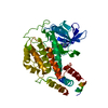
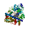
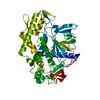
cholesterol 7alpha-monooxygenase / 24-hydroxycholesterol 7alpha-hydroxylase / cholesterol 7-alpha-monooxygenase activity / 24S-hydroxycholesterol 7-alpha-hydroxylase activity / bile acid biosynthetic process / sterol metabolic process / negative regulation of collagen biosynthetic process / regulation of bile acid biosynthetic process / cellular response to cholesterol / negative regulation of fatty acid biosynthetic process ...cholesterol 7alpha-monooxygenase / 24-hydroxycholesterol 7alpha-hydroxylase / cholesterol 7-alpha-monooxygenase activity / 24S-hydroxycholesterol 7-alpha-hydroxylase activity / bile acid biosynthetic process / sterol metabolic process / negative regulation of collagen biosynthetic process / regulation of bile acid biosynthetic process / cellular response to cholesterol / negative regulation of fatty acid biosynthetic process / cholesterol catabolic process / Synthesis of bile acids and bile salts / intracellular membrane-bounded organelle / Synthesis of bile acids and bile salts via 27-hydroxycholesterol / Endogenous sterols / Synthesis of bile acids and bile salts via 7alpha-hydroxycholesterol / cholesterol homeostasis / PPARA activates gene expression / cellular response to glucose stimulus / positive regulation of cholesterol biosynthetic process / response to ethanol / iron ion binding / heme binding / endoplasmic reticulum membrane Similarity search - Function
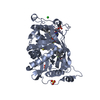
Resolution: 2.75→30 Å / Cor.coef. Fo:Fc: 0.9453 / Cor.coef. Fo:Fc free: 0.9177 / Occupancy max: 1 / Occupancy min: 0.01 / Cross valid method: THROUGHOUT / σ(F): 0 Details: Cholestenone restraints are based on PDB entry 2X5W and were optimized on the prodrg server. Solvent atoms were added based on peaks in the Fo-Fc map. Non-crystallographic symmetry in the ...Details: Cholestenone restraints are based on PDB entry 2X5W and were optimized on the prodrg server. Solvent atoms were added based on peaks in the Fo-Fc map. Non-crystallographic symmetry in the peak positions was considered in the rejection and addition of some sites. Solvent atoms were not included in refinement and structure factor calculation. Some of the assigned water positions likely represent other molecule/atom/ion types. REFMAC, Phenix, coot, and the molprobity server were also used.
In the structure databanks used in Yorodumi, some data are registered as the other names, "COVID-19 virus" and "2019-nCoV". Here are the details of the virus and the list of structure data.
Jan 31, 2019. EMDB accession codes are about to change! (news from PDBe EMDB page)
EMDB accession codes are about to change! (news from PDBe EMDB page)
The allocation of 4 digits for EMDB accession codes will soon come to an end. Whilst these codes will remain in use, new EMDB accession codes will include an additional digit and will expand incrementally as the available range of codes is exhausted. The current 4-digit format prefixed with “EMD-” (i.e. EMD-XXXX) will advance to a 5-digit format (i.e. EMD-XXXXX), and so on. It is currently estimated that the 4-digit codes will be depleted around Spring 2019, at which point the 5-digit format will come into force.
The EM Navigator/Yorodumi systems omit the EMD- prefix.
Related info.:Q: What is EMD? / ID/Accession-code notation in Yorodumi/EM Navigator
Yorodumi is a browser for structure data from EMDB, PDB, SASBDB, etc.
This page is also the successor to EM Navigator detail page, and also detail information page/front-end page for Omokage search.
The word "yorodu" (or yorozu) is an old Japanese word meaning "ten thousand". "mi" (miru) is to see.
Related info.:EMDB / PDB / SASBDB / Comparison of 3 databanks / Yorodumi Search / Aug 31, 2016. New EM Navigator & Yorodumi / Yorodumi Papers / Jmol/JSmol / Function and homology information / Changes in new EM Navigator and Yorodumi
 Movie
Movie Controller

 Yorodumi
 Open data
 Basic information
 Components
 Keywords
 Function and homology information
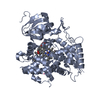
 Homo sapiens (human)
 X-RAY DIFFRACTION /
  Authors
 Citation
 Structure visualization
 Downloads & links
 Other downloads
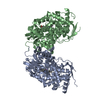


 PDBj
PDBj


 Assembly
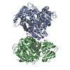


 Mass: 616.487 Da / Num. of mol.: 2 / Source method: obtained synthetically / Formula: C34H32FeN4O4
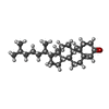
 Mass: 384.638 Da / Num. of mol.: 2 / Source method: obtained synthetically / Formula: C27H44O
 Num. of mol.: 8 / Source method: obtained synthetically
 Mass: 18.015 Da / Num. of mol.: 36 / Source method: isolated from a natural source / Formula: H2O
 Sample preparation
 / Beamline: 23-ID-B / Wavelength: 0.97934
  Processing