 Movie
Movie Controller

[English] 日本語
 Yorodumi
- PDB-3skr: Crystal structure of the 2'- Deoxyguanosine riboswitch bound to 2... -
+ Open data

- Basic information
| Entry | Database: PDB / ID: 3skr | ||||||
|---|---|---|---|---|---|---|---|
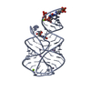
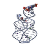
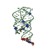
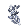
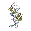
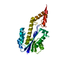
| Title | Crystal structure of the 2'- Deoxyguanosine riboswitch bound to 2'- Deoxyguanosine, cobalt Hexammine soak | ||||||
 Components Components | RNA (66-MER) | ||||||
 Keywords Keywords | RNA / three-way junction / riboswitch / deoxyguanosine | ||||||
| Function / homology | 2'-DEOXY-GUANOSINE / COBALT HEXAMMINE(III) / RNA / RNA (> 10) Function and homology information Function and homology information | ||||||
| Method |  X-RAY DIFFRACTION / SYNCHROTRON / Phaser / Resolution: 3.1 Å X-RAY DIFFRACTION / SYNCHROTRON / Phaser / Resolution: 3.1 Å | ||||||
 Authors Authors | Pikovskaya, O. / Polonskaia, A. / Patel, D.J. / Serganov, A. | ||||||
 Citation Citation | Journal: Nat.Chem.Biol. / Year: 2011 Title: Structural principles of nucleoside selectivity in a 2'-deoxyguanosine riboswitch. Authors: Pikovskaya, O. / Polonskaia, A. / Patel, D.J. / Serganov, A. | ||||||
| History |
|
- Structure visualization
| Structure viewer | Molecule: MolmilJmol/JSmol |
|---|
- Downloads & links
-Download
| PDBx/mmCIF format | 3skr.cif.gz | 83.3 KB | Display | PDBx/mmCIF format |
|---|---|---|---|---|
| PDB format | pdb3skr.ent.gz | 62.6 KB | Display | PDB format |
| PDBx/mmJSON format | 3skr.json.gz | Tree view | PDBx/mmJSON format | |
| Others |  Other downloads Other downloads |
-Validation report
| Summary document | 3skr_validation.pdf.gz | 1007.8 KB | Display | wwPDB validaton report |
|---|---|---|---|---|
| Full document | 3skr_full_validation.pdf.gz | 1017.4 KB | Display | |
| Data in XML | 3skr_validation.xml.gz | 8.1 KB | Display | |
| Data in CIF | 3skr_validation.cif.gz | 10.4 KB | Display | |
| Arichive directory | https://data.pdbj.org/pub/pdb/validation_reports/sk/3skrftp://data.pdbj.org/pub/pdb/validation_reports/sk/3skr | HTTPS FTP |
-Related structure data
| Related structure data |  3skiC 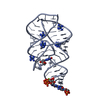 3sklC  3sktC  3skwC  3skzC  3slmC 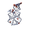 3slqC C: citing same article ( |
|---|---|
| Similar structure data |

-Links
 PDBj
PDBj


- Assembly
| Deposited unit | 
| ||||||||
|---|---|---|---|---|---|---|---|---|---|
| 1 | 
| ||||||||
| 2 | 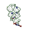
| ||||||||
| Unit cell |
| ||||||||
| Components on special symmetry positions |
|
-Components
| #1: RNA chain | Mass: 21368.484 Da / Num. of mol.: 2 / Source method: obtained synthetically / Details: In vitro transcription #2: Chemical |   Mass: 267.241 Da / Num. of mol.: 2 / Source method: obtained synthetically / Formula: C10H13N5O4 / Details: In vitro transcription Mass: 267.241 Da / Num. of mol.: 2 / Source method: obtained synthetically / Formula: C10H13N5O4 / Details: In vitro transcription#3: Chemical | ChemComp-NCO /   Mass: 161.116 Da / Num. of mol.: 9 / Source method: obtained synthetically / Formula: CoH18N6 Mass: 161.116 Da / Num. of mol.: 9 / Source method: obtained synthetically / Formula: CoH18N6#4: Chemical | ChemComp-MG /   Mass: 24.305 Da / Num. of mol.: 4 / Source method: obtained synthetically / Formula: Mg Mass: 24.305 Da / Num. of mol.: 4 / Source method: obtained synthetically / Formula: Mg#5: Water | ChemComp-HOH / |  Mass: 18.015 Da / Num. of mol.: 18 / Source method: isolated from a natural source / Formula: H2O Mass: 18.015 Da / Num. of mol.: 18 / Source method: isolated from a natural source / Formula: H2O |
|---|
-Experimental details
-Experiment
| Experiment | Method: X-RAY DIFFRACTION / Number of used crystals: 1 |
|---|
- Sample preparation
| Crystal | Density Matthews: 2.23 Å3/Da / Density % sol: 44.94 % |
|---|---|
| Crystal grow | Temperature: 293 K / Method: vapor diffusion, hanging drop / pH: 7.5 Details: 0.05 M Na-HEPES, pH 7.5, ~40 % (v/v) pentaerythritol propoxylate, 0.2 M KCl and 2.0 mM spermidine, VAPOR DIFFUSION, HANGING DROP, temperature 293K |
-Data collection
| Diffraction | Mean temperature: 100 K |
|---|---|
| Diffraction source | Source: SYNCHROTRON / Site: NSLS  / Beamline: X29A / Wavelength: 1.45 Å / Beamline: X29A / Wavelength: 1.45 Å |
| Detector | Type: ADSC QUANTUM 315 / Detector: CCD / Date: Apr 27, 2010 |
| Radiation | Protocol: SINGLE WAVELENGTH / Monochromatic (M) / Laue (L): M / Scattering type: x-ray |
| Radiation wavelength | Wavelength: 1.45 Å / Relative weight: 1 |
| Reflection | Resolution: 3.1→20 Å / Num. all: 7273 / Num. obs: 7041 / % possible obs: 96.8 % / Observed criterion σ(I): 3 |
| Reflection shell | Resolution: 3.1→3.21 Å / Redundancy: 4 % / Rmerge(I) obs: 0.497 / Mean I/σ(I) obs: 3.9 / Num. unique all: 701 / % possible all: 98.2 |
- Processing
| Software |
| |||||||||||||||||||||||||||||||||||||||||||||
|---|---|---|---|---|---|---|---|---|---|---|---|---|---|---|---|---|---|---|---|---|---|---|---|---|---|---|---|---|---|---|---|---|---|---|---|---|---|---|---|---|---|---|---|---|---|---|
| Refinement | Method to determine structure: Phaser / Resolution: 3.1→20 Å / Cor.coef. Fo:Fc: 0.931 / Cor.coef. Fo:Fc free: 0.894 / SU ML: 0.626 / Cross valid method: THROUGHOUT / ESU R Free: 0.666 / Stereochemistry target values: MAXIMUM LIKELIHOOD / Details: HYDROGENS HAVE BEEN ADDED IN THE RIDING POSITIONS
| |||||||||||||||||||||||||||||||||||||||||||||
| Solvent computation | Ion probe radii: 0.8 Å / Shrinkage radii: 0 Å / VDW probe radii: 1.2 Å / Solvent model: MASK | |||||||||||||||||||||||||||||||||||||||||||||
| Refinement step | Cycle: LAST / Resolution: 3.1→20 Å
| |||||||||||||||||||||||||||||||||||||||||||||
| Refine LS restraints |
| |||||||||||||||||||||||||||||||||||||||||||||
| LS refinement shell | Resolution: 3.1→3.179 Å / Total num. of bins used: 20
|
