 ムービー
ムービー コントローラー


+ データを開く

- 基本情報
基本情報
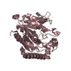
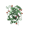
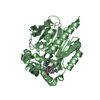
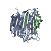
| 登録情報 | データベース: PDB / ID: 3sk0 | ||||||
|---|---|---|---|---|---|---|---|
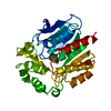
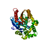
| タイトル | structure of Rhodococcus rhodochrous haloalkane dehalogenase DhaA mutant DhaA12 | ||||||
 要素 要素 | Haloalkane dehalogenase | ||||||
 キーワード キーワード | HYDROLASE / catalytic pentad / alpha/beta-Hydrolase Fold / halide binding / hydrolytic dehalogenation | ||||||
| 機能・相同性 |  機能・相同性情報 機能・相同性情報haloalkane dehalogenase / haloalkane dehalogenase activity / response to toxic substance / membrane 類似検索 - 分子機能 | ||||||
| 生物種 |  Rhodococcus rhodochrous (バクテリア) Rhodococcus rhodochrous (バクテリア) | ||||||
| 手法 |  X線回折 / シンクロトロン / 分子置換 / 解像度: 1.78 Å X線回折 / シンクロトロン / 分子置換 / 解像度: 1.78 Å | ||||||
 データ登録者 データ登録者 | Lahoda, M. / Stsiapanava, A. / Mesters, J. / Koudelakova, T. / Damborsky, J. / Kuta-Smatanova, I. | ||||||
 引用 引用 | ジャーナル: Nat.Chem.Biol. / 年: 2014 タイトル: Dynamics and hydration explain failed functional transformation in dehalogenase design. 著者: Sykora, J. / Brezovsky, J. / Koudelakova, T. / Lahoda, M. / Fortova, A. / Chernovets, T. / Chaloupkova, R. / Stepankova, V. / Prokop, Z. / Smatanova, I.K. / Hof, M. / Damborsky, J. #1: ジャーナル: To Be Publishedタイトル: Crystal structure of haloalkane dehalogenase mutant DhaA12 from Gram-positive bacterium Rhodococcus rhodochrous NCIMB13064 著者: Lahoda, M. / Koudelakova, T. / Stsiapanava, A. / Mesters, J. / Damborsky, J. / Kuta-Smatanova, I. | ||||||
| 履歴 |
|
- 構造の表示
構造の表示
| 構造ビューア | 分子: MolmilJmol/JSmol |
|---|
- ダウンロードとリンク
-ダウンロード
| PDBx/mmCIF形式 | 3sk0.cif.gz | 82.9 KB | 表示 | PDBx/mmCIF形式 |
|---|---|---|---|---|
| PDB形式 | pdb3sk0.ent.gz | 60.1 KB | 表示 | PDB形式 |
| PDBx/mmJSON形式 | 3sk0.json.gz | ツリー表示 | PDBx/mmJSON形式 | |
| その他 |  その他のダウンロード その他のダウンロード |
-検証レポート
| アーカイブディレクトリ | https://data.pdbj.org/pub/pdb/validation_reports/sk/3sk0ftp://data.pdbj.org/pub/pdb/validation_reports/sk/3sk0 | HTTPS FTP |
|---|
-関連構造データ
| 関連構造データ | 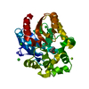 3fbwS S: 精密化の開始モデル |
|---|---|
| 類似構造データ |


-リンク
 PDBj
PDBj


- 集合体
集合体
| 登録構造単位 | 
| ||||||||
|---|---|---|---|---|---|---|---|---|---|
| 1 |
| ||||||||
| 単位格子 |
|
-要素
| #1: タンパク質 | 分子量: 35397.043 Da / 分子数: 1 変異: W138F, 139HHTEVAEEQDH149 insertion, P150A, F152A, G179R, A180V, K183G, C184G, V253A 由来タイプ: 組換発現 由来: (組換発現) Rhodococcus rhodochrous (バクテリア)遺伝子: dhaA / プラスミド: pAQN / 発現宿主: |
|---|---|
| #2: 化合物 | ChemComp-CL /   分子量: 35.453 Da / 分子数: 1 / 由来タイプ: 合成 / 式: Cl 分子量: 35.453 Da / 分子数: 1 / 由来タイプ: 合成 / 式: Cl |
| #3: 水 | ChemComp-HOH /  分子量: 18.015 Da / 分子数: 270 / 由来タイプ: 天然 / 式: H2O 分子量: 18.015 Da / 分子数: 270 / 由来タイプ: 天然 / 式: H2O |
-実験情報
-実験
| 実験 | 手法: X線回折 / 使用した結晶の数: 1 |
|---|
- 試料調製
試料調製
| 結晶 | マシュー密度: 2.11 Å3/Da / 溶媒含有率: 41.74 % |
|---|---|
| 結晶化 | 温度: 277 K / 手法: 蒸気拡散法, シッティングドロップ法 / pH: 6.1 詳細: 100 mM MES sodium salt, 20% peg 4000, pH 6.1, VAPOR DIFFUSION, SITTING DROP, temperature 277K |
-データ収集
| 回折 | 平均測定温度: 100 K |
|---|---|
| 放射光源 | 由来: シンクロトロン / サイト: BESSY  / ビームライン: 14.1 / 波長: 0.918 Å / ビームライン: 14.1 / 波長: 0.918 Å |
| 検出器 | タイプ: MARMOSAIC 225 mm CCD / 検出器: CCD / 日付: 2009年11月24日 |
| 放射 | モノクロメーター: Si 111 / プロトコル: SINGLE WAVELENGTH / 単色(M)・ラウエ(L): M / 散乱光タイプ: x-ray |
| 放射波長 | 波長: 0.918 Å / 相対比: 1 |
| 反射 | 解像度: 1.78→10 Å / Num. obs: 34658 / % possible obs: 100 % / Observed criterion σ(F): 0 / Observed criterion σ(I): 0 / 冗長度: 4.1 % / Biso Wilson estimate: 22.7 Å2 / Rmerge(I) obs: 0.084 / Rsym value: 0.084 / Net I/σ(I): 14.4 |
| 反射 シェル | 解像度: 1.78→1.88 Å / 冗長度: 4.1 % / Rmerge(I) obs: 0.37 / Mean I/σ(I) obs: 4.2 / Num. unique all: 4241 / Rsym value: 0.37 / % possible all: 99.9 |
- 解析
解析
| ソフトウェア |
| |||||||||||||||||||||||||||||||||
|---|---|---|---|---|---|---|---|---|---|---|---|---|---|---|---|---|---|---|---|---|---|---|---|---|---|---|---|---|---|---|---|---|---|---|
| 精密化 | 構造決定の手法: 分子置換 開始モデル: PDB ENTRY 3FBW 解像度: 1.78→10 Å / Num. parameters: 11235 / Num. restraintsaints: 10651 / Isotropic thermal model: isotropic / 交差検証法: FREE R / σ(F): 0 / σ(I): 0 / 立体化学のターゲット値: ENGH AND HUBER 詳細: CONJUGATE GRADIENT LEAST SQUARES REFINEMENT IN SHELXL 97
| |||||||||||||||||||||||||||||||||
| 原子変位パラメータ | Biso mean: 17.1 Å2 | |||||||||||||||||||||||||||||||||
| Refine analyze | Luzzati coordinate error obs: 0.188 Å / Luzzati d res low obs: 6.67 Å / Num. disordered residues: 15 / Occupancy sum hydrogen: 2288 / Occupancy sum non hydrogen: 2751 | |||||||||||||||||||||||||||||||||
| 精密化ステップ | サイクル: LAST / 解像度: 1.78→10 Å
| |||||||||||||||||||||||||||||||||
| 拘束条件 |
| |||||||||||||||||||||||||||||||||
| LS精密化 シェル | 解像度: 1.78→1.88 Å
|
