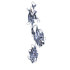
Mass: 33365.621 Da / Num. of mol.: 1 Fragment: 3 N-terminal Immunoglobulin Domains (UNP residues 1-304) Source method: isolated from a genetically manipulated source Details: This structure represents the first three of four domains of Hoc from the bacteriophage RB49. Source: (gene. exp.) Enterobacteria phage RB49 (virus) / Gene: hoc / Production host: Escherichia coli (E. coli) / References: UniProt: Q7Y442
Resolution: 1.95→1.98 Å / Redundancy: 4.7 % / Mean I/σ(I) obs: 4.1 / Rsym value: 0.28 / % possible all: 15
-
Processing
Software
Name
Version
Classification
Blu-Ice
datacollection
PHENIX
Autosol
modelbuilding
SOLVE
phasing
PHENIX
(phenix.refine: 1.7_650)
refinement
HKL-2000
datareduction
HKL-2000
datascaling
PHENIX
Autosol
phasing
Refinement
Method to determine structure: SIRAS, 3-wavelength data from a Pt derivative Resolution: 1.951→27.691 Å / SU ML: 0.32 / σ(F): 0 / σ(I): 0 / Phase error: 36.81 / Stereochemistry target values: ML Details: Diffraction data were anisotropic. Resolution of the dataset along the three principle mutually perpendicular directions determined by the vectors a*/|a*| + c*/|c*|; a*/|a*| - c*/|c*|, and ...Details: Diffraction data were anisotropic. Resolution of the dataset along the three principle mutually perpendicular directions determined by the vectors a*/|a*| + c*/|c*|; a*/|a*| - c*/|c*|, and b* was 1.95, 2.8 and 2.7 A, respectively (resolution at which average I/sigma = 2). The data were truncated using an ellipsoid which had its principle axes along these vectors. All reflections which were outside the ellipsoid and had I/sigma(I) < 2.0 were rejected. The structure factors were modified by applying an anisotropic scaling to scale up reflections located in the weak directions of reciprocal space. The components of the B-factor scaling tensor corresponding to the principle axes of the ellipsoid were 0, -60, -35 A2, respectively.
Rfactor
Num. reflection
% reflection
Selection details
Rfree
0.2673
1766
9.76 %
RANDOM
Rwork
0.2181
-
-
-
all
0.2231
35571
-
-
obs
0.2231
18095
50.87 %
-
Solvent computation
Shrinkage radii: 1.25 Å / VDW probe radii: 1.3 Å / Solvent model: FLAT BULK SOLVENT MODEL / Bsol: 45.254 Å2 / ksol: 0.336 e/Å3
Displacement parameters
Baniso -1
Baniso -2
Baniso -3
1-
-6.7957 Å2
0 Å2
-5.708 Å2
2-
-
-9.2991 Å2
0 Å2
3-
-
-
-4.9752 Å2
Refinement step
Cycle: LAST / Resolution: 1.951→27.691 Å
Protein
Nucleic acid
Ligand
Solvent
Total
Num. atoms
2333
0
1
90
2424
Refine LS restraints
Refine-ID
Type
Dev ideal
Number
X-RAY DIFFRACTION
f_bond_d
0.008
2376
X-RAY DIFFRACTION
f_angle_d
1.175
3238
X-RAY DIFFRACTION
f_dihedral_angle_d
14.301
840
X-RAY DIFFRACTION
f_chiral_restr
0.086
380
X-RAY DIFFRACTION
f_plane_restr
0.005
418
LS refinement shell
Resolution (Å)
Rfactor Rfree
Num. reflection Rfree
Rfactor Rwork
Num. reflection Rwork
Refine-ID
% reflection obs (%)
1.9511-2.0208
0.3074
48
0.2334
396
X-RAY DIFFRACTION
13
2.0208-2.1017
0.3195
44
0.249
522
X-RAY DIFFRACTION
16
2.1017-2.1973
0.3429
75
0.2819
715
X-RAY DIFFRACTION
23
2.1973-2.3131
0.4665
40
0.3999
383
X-RAY DIFFRACTION
12
2.3131-2.4579
0.3176
126
0.309
1161
X-RAY DIFFRACTION
37
2.4579-2.6476
0.374
163
0.3061
1635
X-RAY DIFFRACTION
51
2.6476-2.9138
0.3639
297
0.295
2716
X-RAY DIFFRACTION
85
2.9138-3.3348
0.3075
364
0.2583
3228
X-RAY DIFFRACTION
100
3.3348-4.1991
0.2677
250
0.197
2337
X-RAY DIFFRACTION
72
4.1991-27.6943
0.1929
359
0.1551
3236
X-RAY DIFFRACTION
97
Refinement TLS params.
Method: refined / Refine-ID: X-RAY DIFFRACTION
ID
L11 (°2)
L12 (°2)
L13 (°2)
L22 (°2)
L23 (°2)
L33 (°2)
S11 (Å °)
S12 (Å °)
S13 (Å °)
S21 (Å °)
S22 (Å °)
S23 (Å °)
S31 (Å °)
S32 (Å °)
S33 (Å °)
T11 (Å2)
T12 (Å2)
T13 (Å2)
T22 (Å2)
T23 (Å2)
T33 (Å2)
Origin x (Å)
Origin y (Å)
Origin z (Å)
1
0.5015
-0.0119
-0.2005
0.5788
-0.036
0.283
0.0364
-0.1132
0.0217
-0.0376
0.0185
0.1733
-0.0619
0.0084
0.0301
0.1619
-0.0112
-0.0561
0.0928
0.0577
0.3752
-38.9731
25.5143
-55.2805
2
0.1339
0.0001
0.238
0.3254
-0.1167
0.552
0.0567
-0.3923
-0.1358
-0.0149
0.0421
0.1224
0.0744
0.0486
-0.053
0.0351
0.0001
-0.1571
0.9518
0.2155
0.1568
-12.7528
15.4445
-28.8754
3
0.1359
-0.1349
-0.1597
0.2236
0.0501
0.5663
0.0485
-0.0526
0.0928
0.1086
-0.0069
-0.2658
-0.1236
0.3153
-0.0109
-0.0091
-0.1051
-0.0406
1.3264
-0.0766
0.2474
15.2963
25.1913
-2.0479
Refinement TLS group
ID
Refine-ID
Refine TLS-ID
Selection details
1
X-RAY DIFFRACTION
1
chain 'A' and ((resseq2:89))
2
X-RAY DIFFRACTION
2
chain 'A' and ((resseq90:181))
3
X-RAY DIFFRACTION
3
chain 'A' and ((resseq182:304))
+
About Yorodumi
-
News
-
Feb 9, 2022. New format data for meta-information of EMDB entries
New format data for meta-information of EMDB entries
Version 3 of the EMDB header file is now the official format.
The previous official version 1.9 will be removed from the archive.
In the structure databanks used in Yorodumi, some data are registered as the other names, "COVID-19 virus" and "2019-nCoV". Here are the details of the virus and the list of structure data.
Jan 31, 2019. EMDB accession codes are about to change! (news from PDBe EMDB page)
EMDB accession codes are about to change! (news from PDBe EMDB page)
The allocation of 4 digits for EMDB accession codes will soon come to an end. Whilst these codes will remain in use, new EMDB accession codes will include an additional digit and will expand incrementally as the available range of codes is exhausted. The current 4-digit format prefixed with “EMD-” (i.e. EMD-XXXX) will advance to a 5-digit format (i.e. EMD-XXXXX), and so on. It is currently estimated that the 4-digit codes will be depleted around Spring 2019, at which point the 5-digit format will come into force.
The EM Navigator/Yorodumi systems omit the EMD- prefix.
Related info.:Q: What is EMD? / ID/Accession-code notation in Yorodumi/EM Navigator
Yorodumi is a browser for structure data from EMDB, PDB, SASBDB, etc.
This page is also the successor to EM Navigator detail page, and also detail information page/front-end page for Omokage search.
The word "yorodu" (or yorozu) is an old Japanese word meaning "ten thousand". "mi" (miru) is to see.
Related info.:EMDB / PDB / SASBDB / Comparison of 3 databanks / Yorodumi Search / Aug 31, 2016. New EM Navigator & Yorodumi / Yorodumi Papers / Jmol/JSmol / Function and homology information / Changes in new EM Navigator and Yorodumi
 Movie
Movie Controller

 Yorodumi
 Open data
 Basic information
 Components
 Keywords
 Function and homology information
 Enterobacteria phage RB49 (virus)
 X-RAY DIFFRACTION /
  Authors
 Citation
 Structure visualization
 Downloads & links
 Other downloads
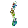


 PDBj
PDBj Assembly
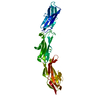


 Mass: 24.305 Da / Num. of mol.: 1 / Source method: obtained synthetically / Formula: Mg
 Mass: 18.015 Da / Num. of mol.: 90 / Source method: isolated from a natural source / Formula: H2O
 Sample preparation
 / Beamline: 23-ID-D / Wavelength: 0.9795 Å
 Processing