 Movie
Movie Controller


+ Open data

- Basic information
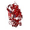
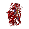
| Entry | Database: PDB / ID: 6u7n | ||||||
|---|---|---|---|---|---|---|---|
| Title | Crystal structure of neurotrimin (NTM) | ||||||
 Components Components | Neurotrimin | ||||||
 Keywords Keywords | CELL ADHESION / synaptic organizer / IgLON / Ig domain-containing | ||||||
| Function / homology |  Function and homology information Function and homology informationneuron recognition / Post-translational modification: synthesis of GPI-anchored proteins / side of membrane / cell adhesion / extracellular region / plasma membrane Similarity search - Function | ||||||
| Biological species |  Homo sapiens (human) Homo sapiens (human) | ||||||
| Method |  X-RAY DIFFRACTION / SYNCHROTRON / MOLECULAR REPLACEMENT / Resolution: 3.321 Å X-RAY DIFFRACTION / SYNCHROTRON / MOLECULAR REPLACEMENT / Resolution: 3.321 Å | ||||||
 Authors Authors | Machius, M. / Venkannagari, H. / Misra, A. / Rudenko, G. / Rush, S. | ||||||
| Funding support |  United States, 1items United States, 1items
| ||||||
 Citation Citation | Journal: J.Mol.Biol. / Year: 2020 Title: Highly Conserved Molecular Features in IgLONs Contrast Their Distinct Structural and Biological Outcomes. Authors: Venkannagari, H. / Kasper, J.M. / Misra, A. / Rush, S.A. / Fan, S. / Lee, H. / Sun, H. / Seshadrinathan, S. / Machius, M. / Hommel, J.D. / Rudenko, G. | ||||||
| History |
|
- Structure visualization
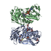
| Structure viewer | Molecule: MolmilJmol/JSmol |
|---|
- Downloads & links
-Download
| PDBx/mmCIF format | 6u7n.cif.gz | 170.1 KB | Display | PDBx/mmCIF format |
|---|---|---|---|---|
| PDB format | pdb6u7n.ent.gz | 135.3 KB | Display | PDB format |
| PDBx/mmJSON format | 6u7n.json.gz | Tree view | PDBx/mmJSON format | |
| Others |  Other downloads Other downloads |
-Validation report
| Arichive directory | https://data.pdbj.org/pub/pdb/validation_reports/u7/6u7nftp://data.pdbj.org/pub/pdb/validation_reports/u7/6u7n | HTTPS FTP |
|---|
-Related structure data
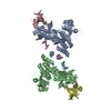
| Related structure data |  6u6tC 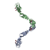 5uv6S S: Starting model for refinement C: citing same article ( |
|---|---|
| Similar structure data |
-Links
 PDBj
PDBj


- Assembly
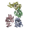
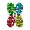
| Deposited unit | 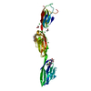
| ||||||||
|---|---|---|---|---|---|---|---|---|---|
| 1 | 
| ||||||||
| Unit cell |
|
-Components
| #1: Protein | Mass: 34812.332 Da / Num. of mol.: 1 Source method: isolated from a genetically manipulated source Source: (gene. exp.) Homo sapiens (human) / Gene: NTM, IGLON2, NT, UNQ297/PRO337 / Plasmid: pFastbac / Production host:  Baculovirus expression vector pFastBac1-HM / References: UniProt: Q9P121 Baculovirus expression vector pFastBac1-HM / References: UniProt: Q9P121 | ||||||
|---|---|---|---|---|---|---|---|
| #2: Polysaccharide | beta-D-mannopyranose-(1-4)-2-acetamido-2-deoxy-beta-D-glucopyranose-(1-4)-2-acetamido-2-deoxy-beta- ...beta-D-mannopyranose-(1-4)-2-acetamido-2-deoxy-beta-D-glucopyranose-(1-4)-2-acetamido-2-deoxy-beta-D-glucopyranose Source method: isolated from a genetically manipulated source | ||||||
| #3: Polysaccharide | alpha-L-fucopyranose-(1-6)-2-acetamido-2-deoxy-beta-D-glucopyranose Source method: isolated from a genetically manipulated source | ||||||
| #4: Chemical |   Mass: 62.068 Da / Num. of mol.: 3 Mass: 62.068 Da / Num. of mol.: 3Source method: isolated from a genetically manipulated source Formula: C2H6O2 #5: Chemical | ChemComp-CL /   Mass: 35.453 Da / Num. of mol.: 8 Mass: 35.453 Da / Num. of mol.: 8Source method: isolated from a genetically manipulated source Formula: Cl Has ligand of interest | N | Has protein modification | Y | |
-Experimental details
-Experiment
| Experiment | Method: X-RAY DIFFRACTION / Number of used crystals: 1 |
|---|
- Sample preparation
| Crystal grow | Temperature: 293.15 K / Method: vapor diffusion, hanging drop / pH: 4.5 Details: Protein: 5.4 mg/ml NTM in 10 mM HEPES, pH 8.0, 150 mM sodium chloride Reservoir Solution: 2.5 M NaCl, 100 mM sodium acetate pH 4.5, 200 mM lithium sulfate Crystallization Drop: 2 micro- ...Details: Protein: 5.4 mg/ml NTM in 10 mM HEPES, pH 8.0, 150 mM sodium chloride Reservoir Solution: 2.5 M NaCl, 100 mM sodium acetate pH 4.5, 200 mM lithium sulfate Crystallization Drop: 2 micro-Liters NTM + 2 micro-Liters Reservoir Solution Single crystals grew to a size of ~250 x 150 micro-meters within 7-10 days. Crystals harvested from the crystallization drops were cryo-protected in reservoir solution containing 20% (v/v) ethylene glycol and then flash cooled by plunging into liquid nitrogen |
|---|
-Data collection
| Diffraction | Mean temperature: 100 K / Serial crystal experiment: N | |||||||||||||||||||||||||||||||||||||||||||||||||||||||||||||||||||||||||||||||||||||||||||||||||||||||||||||||||||||||||||||||||||||||||||||||||||||||||||||||||||||||||||||||||||||||||||||
|---|---|---|---|---|---|---|---|---|---|---|---|---|---|---|---|---|---|---|---|---|---|---|---|---|---|---|---|---|---|---|---|---|---|---|---|---|---|---|---|---|---|---|---|---|---|---|---|---|---|---|---|---|---|---|---|---|---|---|---|---|---|---|---|---|---|---|---|---|---|---|---|---|---|---|---|---|---|---|---|---|---|---|---|---|---|---|---|---|---|---|---|---|---|---|---|---|---|---|---|---|---|---|---|---|---|---|---|---|---|---|---|---|---|---|---|---|---|---|---|---|---|---|---|---|---|---|---|---|---|---|---|---|---|---|---|---|---|---|---|---|---|---|---|---|---|---|---|---|---|---|---|---|---|---|---|---|---|---|---|---|---|---|---|---|---|---|---|---|---|---|---|---|---|---|---|---|---|---|---|---|---|---|---|---|---|---|---|---|---|---|
| Diffraction source | Source: SYNCHROTRON / Site: ALS / Beamline: 8.2.1 / Wavelength: 0.99999 Å | |||||||||||||||||||||||||||||||||||||||||||||||||||||||||||||||||||||||||||||||||||||||||||||||||||||||||||||||||||||||||||||||||||||||||||||||||||||||||||||||||||||||||||||||||||||||||||||
| Detector | Type: ADSC QUANTUM 315 / Detector: CCD / Date: May 31, 2018 | |||||||||||||||||||||||||||||||||||||||||||||||||||||||||||||||||||||||||||||||||||||||||||||||||||||||||||||||||||||||||||||||||||||||||||||||||||||||||||||||||||||||||||||||||||||||||||||
| Radiation | Protocol: SINGLE WAVELENGTH / Monochromatic (M) / Laue (L): M / Scattering type: x-ray | |||||||||||||||||||||||||||||||||||||||||||||||||||||||||||||||||||||||||||||||||||||||||||||||||||||||||||||||||||||||||||||||||||||||||||||||||||||||||||||||||||||||||||||||||||||||||||||
| Radiation wavelength | Wavelength: 0.99999 Å / Relative weight: 1 | |||||||||||||||||||||||||||||||||||||||||||||||||||||||||||||||||||||||||||||||||||||||||||||||||||||||||||||||||||||||||||||||||||||||||||||||||||||||||||||||||||||||||||||||||||||||||||||
| Reflection | Resolution: 3.32→46.431 Å / Num. obs: 18288 / % possible obs: 99.4 % / Redundancy: 4.6 % / Rmerge(I) obs: 0.062 / Rpim(I) all: 0.032 / Rrim(I) all: 0.07 / Χ2: 0.845 / Net I/σ(I): 13 | |||||||||||||||||||||||||||||||||||||||||||||||||||||||||||||||||||||||||||||||||||||||||||||||||||||||||||||||||||||||||||||||||||||||||||||||||||||||||||||||||||||||||||||||||||||||||||||
| Reflection shell | Diffraction-ID: 1
|
- Processing
| Software |
| ||||||||||||||||||||||||||||||||||||||||
|---|---|---|---|---|---|---|---|---|---|---|---|---|---|---|---|---|---|---|---|---|---|---|---|---|---|---|---|---|---|---|---|---|---|---|---|---|---|---|---|---|---|
| Refinement | Method to determine structure: MOLECULAR REPLACEMENT Starting model: 5UV6 Resolution: 3.321→46.43 Å / SU ML: 0.38 / Cross valid method: THROUGHOUT / σ(F): 1.36 / Phase error: 26.56
| ||||||||||||||||||||||||||||||||||||||||
| Solvent computation | Shrinkage radii: 0.9 Å / VDW probe radii: 1.11 Å | ||||||||||||||||||||||||||||||||||||||||
| Displacement parameters | Biso max: 234.26 Å2 / Biso mean: 79.78 Å2 / Biso min: 27.76 Å2 | ||||||||||||||||||||||||||||||||||||||||
| Refinement step | Cycle: final / Resolution: 3.321→46.43 Å
| ||||||||||||||||||||||||||||||||||||||||
| LS refinement shell | Refine-ID: X-RAY DIFFRACTION / Rfactor Rfree error: 0
| ||||||||||||||||||||||||||||||||||||||||
| Refinement TLS params. | Method: refined / Origin x: 5.2396 Å / Origin y: -38.7208 Å / Origin z: 4.6635 Å
| ||||||||||||||||||||||||||||||||||||||||
| Refinement TLS group |
|
