- PDB-3shf: Crystal structure of the R265S mutant of full-length murine Apaf-1 -
+
Open data
ID or keywords:
Loading...
-
Basic information
Entry
Database: PDB / ID: 3shf
Title
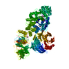
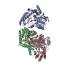
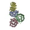
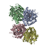
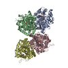
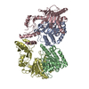
Crystal structure of the R265S mutant of full-length murine Apaf-1
Components
Apoptotic peptidase activating factor 1
Keywords
APOPTOSIS / tandem beta-propeller / cytochrome c / adenine nucleotide / procaspase-9 / cytosol
Function / homology
Function and homology information
Formation of apoptosome / Activation of caspases through apoptosome-mediated cleavage / Regulation of the apoptosome activity / regulation of apoptotic DNA fragmentation / apoptosome / cysteine-type endopeptidase activator activity involved in apoptotic process / forebrain development / intrinsic apoptotic signaling pathway in response to endoplasmic reticulum stress / cellular response to transforming growth factor beta stimulus / cardiac muscle cell apoptotic process ...Formation of apoptosome / Activation of caspases through apoptosome-mediated cleavage / Regulation of the apoptosome activity / regulation of apoptotic DNA fragmentation / apoptosome / cysteine-type endopeptidase activator activity involved in apoptotic process / forebrain development / intrinsic apoptotic signaling pathway in response to endoplasmic reticulum stress / cellular response to transforming growth factor beta stimulus / cardiac muscle cell apoptotic process / heat shock protein binding / Neutrophil degranulation / response to nutrient / intrinsic apoptotic signaling pathway / positive regulation of apoptotic signaling pathway / kidney development / neural tube closure / ADP binding / brain development / neuron apoptotic process / response to hypoxia / cell differentiation / positive regulation of apoptotic process / protein-containing complex / ATP binding / identical protein binding / nucleus / cytoplasm / cytosol Similarity search - Function
In the structure databanks used in Yorodumi, some data are registered as the other names, "COVID-19 virus" and "2019-nCoV". Here are the details of the virus and the list of structure data.
Jan 31, 2019. EMDB accession codes are about to change! (news from PDBe EMDB page)
EMDB accession codes are about to change! (news from PDBe EMDB page)
The allocation of 4 digits for EMDB accession codes will soon come to an end. Whilst these codes will remain in use, new EMDB accession codes will include an additional digit and will expand incrementally as the available range of codes is exhausted. The current 4-digit format prefixed with “EMD-” (i.e. EMD-XXXX) will advance to a 5-digit format (i.e. EMD-XXXXX), and so on. It is currently estimated that the 4-digit codes will be depleted around Spring 2019, at which point the 5-digit format will come into force.
The EM Navigator/Yorodumi systems omit the EMD- prefix.
Related info.:Q: What is EMD? / ID/Accession-code notation in Yorodumi/EM Navigator
Yorodumi is a browser for structure data from EMDB, PDB, SASBDB, etc.
This page is also the successor to EM Navigator detail page, and also detail information page/front-end page for Omokage search.
The word "yorodu" (or yorozu) is an old Japanese word meaning "ten thousand". "mi" (miru) is to see.
Related info.:EMDB / PDB / SASBDB / Comparison of 3 databanks / Yorodumi Search / Aug 31, 2016. New EM Navigator & Yorodumi / Yorodumi Papers / Jmol/JSmol / Function and homology information / Changes in new EM Navigator and Yorodumi
 Movie
Movie Controller

 Yorodumi
 Open data
 Basic information
 Components
 Keywords
 Function and homology information
 X-RAY DIFFRACTION /
  Authors
 Citation
 Structure visualization
 Downloads & links
 Other downloads


 PDBj
PDBj


 Assembly
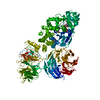

 Spodoptera frugiperda (fall armyworm) / References: UniProt: A2RRK8, UniProt: O88879*PLUS
 Mass: 427.201 Da / Num. of mol.: 1 / Source method: obtained synthetically / Formula: C10H15N5O10P2 / Comment: ADP, energy-carrying molecule*YM
 Mass: 86.089 Da / Num. of mol.: 2 / Source method: obtained synthetically / Formula: C4H6O2
 Mass: 18.015 Da / Num. of mol.: 73 / Source method: isolated from a natural source / Formula: H2O
 Sample preparation
 / Beamline: ID23-2 / Wavelength: 0.8726 Å
 Processing