 Movie
Movie Controller

[English] 日本語
 Yorodumi
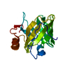
- PDB-3sft: Crystal structure of Thermotoga maritima CheB methylesterase cata... -
+ Open data

- Basic information
| Entry | Database: PDB / ID: 3sft | ||||||
|---|---|---|---|---|---|---|---|
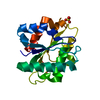
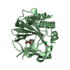
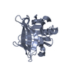
| Title | Crystal structure of Thermotoga maritima CheB methylesterase catalytic domain | ||||||
 Components Components | Chemotaxis response regulator protein-glutamate methylesterase | ||||||
 Keywords Keywords | HYDROLASE / modified doubly-wound/fold / Methylesterase / chemoreceptor | ||||||
| Function / homology |  Function and homology information Function and homology informationprotein-glutamate methylesterase / protein-glutamate methylesterase activity / protein-glutamine glutaminase activity / protein-glutamine glutaminase / phosphorelay response regulator activity / chemotaxis / signal transduction / cytoplasm Similarity search - Function | ||||||
| Biological species |   Thermotoga maritima (bacteria) Thermotoga maritima (bacteria) | ||||||
| Method |  X-RAY DIFFRACTION / SYNCHROTRON / MOLECULAR REPLACEMENT / Resolution: 2.15 Å X-RAY DIFFRACTION / SYNCHROTRON / MOLECULAR REPLACEMENT / Resolution: 2.15 Å | ||||||
 Authors Authors | Park, S.Y. / Crane, B.R. | ||||||
 Citation Citation | Journal: Biochem.Biophys.Res.Commun. / Year: 2011 Title: An insight into the interaction mode between CheB and chemoreceptor from two crystal structures of CheB methylesterase catalytic domain Authors: Cho, K.H. / Crane, B.R. / Park, S.Y. | ||||||
| History |
|
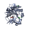
- Structure visualization
| Structure viewer | Molecule: MolmilJmol/JSmol |
|---|
- Downloads & links
-Download
| PDBx/mmCIF format | 3sft.cif.gz | 50.4 KB | Display | PDBx/mmCIF format |
|---|---|---|---|---|
| PDB format | pdb3sft.ent.gz | 35.7 KB | Display | PDB format |
| PDBx/mmJSON format | 3sft.json.gz | Tree view | PDBx/mmJSON format | |
| Others |  Other downloads Other downloads |
-Validation report
| Arichive directory | https://data.pdbj.org/pub/pdb/validation_reports/sf/3sftftp://data.pdbj.org/pub/pdb/validation_reports/sf/3sft | HTTPS FTP |
|---|
-Related structure data
| Related structure data | 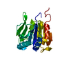 1chdS S: Starting model for refinement |
|---|---|
| Similar structure data |


-Links
 PDBj
PDBj


- Assembly
| Deposited unit | 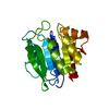
| ||||||||
|---|---|---|---|---|---|---|---|---|---|
| 1 |
| ||||||||
| Unit cell |
|
-Components
| #1: Protein | Mass: 20895.289 Da / Num. of mol.: 1 / Fragment: CheB methylesterase catalytic domain Source method: isolated from a genetically manipulated source Source: (gene. exp.) Thermotoga maritima (bacteria) / Gene: cheB / Plasmid: pET28a / Production host: References: UniProt: Q9WYN9, protein-glutamate methylesterase |
|---|---|
| #2: Water | ChemComp-HOH /  Mass: 18.015 Da / Num. of mol.: 106 / Source method: isolated from a natural source / Formula: H2O Mass: 18.015 Da / Num. of mol.: 106 / Source method: isolated from a natural source / Formula: H2O |
-Experimental details
-Experiment
| Experiment | Method: X-RAY DIFFRACTION / Number of used crystals: 1 |
|---|
- Sample preparation
| Crystal | Density Matthews: 3.27 Å3/Da / Density % sol: 62.33 % |
|---|---|
| Crystal grow | Temperature: 298 K / Method: vapor diffusion, hanging drop / pH: 6.5 Details: 0.2M ammonium sulfate, 0.1M cacodylate, 30%(w/v) PEG 8K, pH 6.5, VAPOR DIFFUSION, HANGING DROP, temperature 298K |
-Data collection
| Diffraction | Mean temperature: 100 K |
|---|---|
| Diffraction source | Source: SYNCHROTRON / Site: NSLS  / Beamline: X25 / Beamline: X25 |
| Detector | Type: ADSC QUANTUM 315 / Detector: CCD / Date: Jan 23, 2004 |
| Radiation | Protocol: SINGLE WAVELENGTH / Monochromatic (M) / Laue (L): M / Scattering type: x-ray |
| Radiation wavelength | Relative weight: 1 |
| Reflection | Resolution: 2.15→30 Å / Num. obs: 13555 |
- Processing
| Software |
| ||||||||||||||||||||
|---|---|---|---|---|---|---|---|---|---|---|---|---|---|---|---|---|---|---|---|---|---|
| Refinement | Method to determine structure: MOLECULAR REPLACEMENT Starting model: 1CHD Resolution: 2.15→30 Å
| ||||||||||||||||||||
| Refinement step | Cycle: LAST / Resolution: 2.15→30 Å
|
