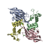
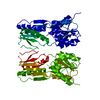
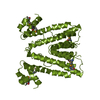
- PDB-3sb3: Crystal structure of an apag protein (PA1934) from pseudomonas ae... -
+
Open data
ID or keywords:
Loading...
-
Basic information
Entry
Database: PDB / ID: 3sb3
Title
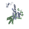
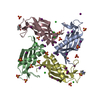
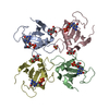
Crystal structure of an apag protein (PA1934) from pseudomonas aeruginosa pao1 at 1.83 A resolution
Components
ApaG protein
Keywords
Structural Genomics / Unknown Function / Joint Center for Structural Genomics / JCSG / Protein Structure Initiative / PSI-BIOLOGY
Function / homology
Protein of unknown function DUF4354 / Protein of unknown function DUF4354 / Domain of unknown function (DUF4354) / Immunoglobulin-like / Sandwich / Mainly Beta / PHOSPHATE ION / DUF4354 family protein
Function and homology information
Biological species
Pseudomonas aeruginosa (bacteria)
Method
X-RAY DIFFRACTION / SYNCHROTRON / MAD / Resolution: 1.83 Å
Mass: 18.015 Da / Num. of mol.: 77 / Source method: isolated from a natural source / Formula: H2O
Has protein modification
Y
Sequence details
THIS CONSTRUCT (RESIDUES 22-126) WAS EXPRESSED WITH A PURIFICATION TAG MGSDKIHHHHHHENLYFQG. THE TAG ...THIS CONSTRUCT (RESIDUES 22-126) WAS EXPRESSED WITH A PURIFICATION TAG MGSDKIHHHHHHENLYFQG. THE TAG WAS REMOVED WITH TEV PROTEASE LEAVING ONLY A GLYCINE (0) FOLLOWED BY THE TARGET SEQUENCE
-
Experimental details
-
Experiment
Experiment
Method: X-RAY DIFFRACTION / Number of used crystals: 1
-
Sample preparation
Crystal
Density Matthews: 2.46 Å3/Da / Density % sol: 50.08 %
Crystal grow
Temperature: 277 K / Method: vapor diffusion, sitting drop / pH: 8.5 Details: 0.2M MgCl2, 30.00% PEG-400, 0.1M TRIS pH 8.5, NANODROP, VAPOR DIFFUSION, SITTING DROP, temperature 277K
Resolution: 1.83→29.552 Å / Num. obs: 10313 / % possible obs: 99.3 % / Observed criterion σ(I): -3 / Redundancy: 7.69 % / Biso Wilson estimate: 27.998 Å2 / Rmerge(I) obs: 0.052 / Net I/σ(I): 18.07
Reflection shell
Diffraction-ID: 1
Resolution (Å)
Highest resolution (Å)
Rmerge(I) obs
Mean I/σ(I) obs
Num. measured obs
Num. unique obs
% possible all
1.83-1.9
0.79
1.86
7786
1910
94.5
1.9-1.97
0.546
2.8
7237
1720
99.8
1.97-2.06
0.324
4.7
7989
1887
99.9
2.06-2.17
0.243
6.3
8144
1929
99.8
2.17-2.31
0.171
8.6
8206
1943
99.8
2.31-2.48
0.132
11.4
7582
1781
99.9
2.48-2.73
0.086
16
8098
1898
100
2.73-3.13
0.048
26.5
8255
1930
99.9
3.13-3.93
0.027
44.3
7935
1867
100
3.93
0.021
56.9
8061
1910
99.4
-
Phasing
Phasing
Method: MAD
-
Processing
Software
Name
Version
Classification
NB
MolProbity
3beta29
modelbuilding
PDB_EXTRACT
3.1
dataextraction
SHELX
phasing
SHARP
phasing
XSCALE
January30, 2009
datascaling
REFMAC
5.5.0110
refinement
XDS
datareduction
SHELXD
phasing
Refinement
Method to determine structure: MAD / Resolution: 1.83→29.552 Å / Cor.coef. Fo:Fc: 0.961 / Cor.coef. Fo:Fc free: 0.942 / Occupancy max: 1 / Occupancy min: 0.5 / SU B: 5.199 / SU ML: 0.081 / Cross valid method: THROUGHOUT / σ(F): 0 / ESU R Free: 0.122 Stereochemistry target values: MAXIMUM LIKELIHOOD WITH PHASES Details: 1. HYDROGENS HAVE BEEN ADDED IN THE RIDING POSITIONS. 2. ATOM RECORD CONTAINS SUM OF TLS AND RESIDUAL B FACTORS. ANISOU RECORD CONTAINS SUM OF TLS AND RESIDUAL U FACTORS. 3 .A MET-INHIBITION ...Details: 1. HYDROGENS HAVE BEEN ADDED IN THE RIDING POSITIONS. 2. ATOM RECORD CONTAINS SUM OF TLS AND RESIDUAL B FACTORS. ANISOU RECORD CONTAINS SUM OF TLS AND RESIDUAL U FACTORS. 3 .A MET-INHIBITION PROTOCOL WAS USED FOR SELENOMETHIONINE INCORPORATION DURING PROTEIN EXPRESSION. THE OCCUPANCY OF THE SE ATOMS IN THE MSE RESIDUES WAS REDUCED TO 0.75 FOR THE REDUCED SCATTERING POWER DUE TO PARTIAL S-MET INCORPORATION 4.SOLVENT MOLECULES WERE EXCLUDED FROM THE ASSIGNMENT OF THE TLS GROUPS. 5. BASED ON FIT TO ELECTRON DENSITY, A PHOSPHATE (PO4) HAS BEEN MODELED AT A SPECIAL POSITION NEAR THE SIDECHAINS OF ARG 124 AND ARG 120. HOWEVER, THIS ASSIGNMENT OF ELECTRON DENSITY TO A PHOSPHATE IS TENTATIVE.
Rfactor
Num. reflection
% reflection
Selection details
Rfree
0.2227
497
4.8 %
RANDOM
Rwork
0.1872
-
-
-
obs
0.1888
10296
99.61 %
-
Solvent computation
Ion probe radii: 0.8 Å / Shrinkage radii: 0.8 Å / VDW probe radii: 1.4 Å / Solvent model: MASK
In the structure databanks used in Yorodumi, some data are registered as the other names, "COVID-19 virus" and "2019-nCoV". Here are the details of the virus and the list of structure data.
Jan 31, 2019. EMDB accession codes are about to change! (news from PDBe EMDB page)
EMDB accession codes are about to change! (news from PDBe EMDB page)
The allocation of 4 digits for EMDB accession codes will soon come to an end. Whilst these codes will remain in use, new EMDB accession codes will include an additional digit and will expand incrementally as the available range of codes is exhausted. The current 4-digit format prefixed with “EMD-” (i.e. EMD-XXXX) will advance to a 5-digit format (i.e. EMD-XXXXX), and so on. It is currently estimated that the 4-digit codes will be depleted around Spring 2019, at which point the 5-digit format will come into force.
The EM Navigator/Yorodumi systems omit the EMD- prefix.
Related info.:Q: What is EMD? / ID/Accession-code notation in Yorodumi/EM Navigator
Yorodumi is a browser for structure data from EMDB, PDB, SASBDB, etc.
This page is also the successor to EM Navigator detail page, and also detail information page/front-end page for Omokage search.
The word "yorodu" (or yorozu) is an old Japanese word meaning "ten thousand". "mi" (miru) is to see.
Related info.:EMDB / PDB / SASBDB / Comparison of 3 databanks / Yorodumi Search / Aug 31, 2016. New EM Navigator & Yorodumi / Yorodumi Papers / Jmol/JSmol / Function and homology information / Changes in new EM Navigator and Yorodumi
 Movie
Movie Controller

 Yorodumi
 Open data
 Basic information
 Components
 Keywords
 Function and homology information
 Pseudomonas aeruginosa (bacteria)
 X-RAY DIFFRACTION /
  Authors
 Citation
 Structure visualization
 Downloads & links
 Other downloads

 PDBj
PDBj Assembly


 Mass: 94.971 Da / Num. of mol.: 1 / Source method: obtained synthetically / Formula: PO4
 Mass: 18.015 Da / Num. of mol.: 77 / Source method: isolated from a natural source / Formula: H2O
 Sample preparation
 / Beamline: BL9-2 / Wavelength: 0.91837,0.97936,0.97920
 Processing