 Movie
Movie Controller


+ Open data

- Basic information
| Entry | Database: PDB / ID: 3sar | ||||||
|---|---|---|---|---|---|---|---|
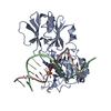
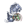
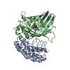
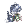
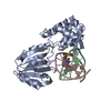
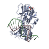
| Title | MUTM Slanted complex 1 | ||||||
 Components Components |
| ||||||
 Keywords Keywords | HYDROLASE/DNA / DNA GLYCOSYLASE / DNA REPAIR / DAMAGE SEARCH / TRANSLOCATION / DISULFIDE CROSSLINKING / DNA DAMAGE / DNA-BINDING / GLYCOSIDASE / HYDROLASE / LYASE / METAL-BINDING / MULTIFUNCTIONAL ENZYME / ZINC-FINGER / HYDROLASE-DNA complex | ||||||
| Function / homology |  Function and homology information Function and homology informationDNA-formamidopyrimidine glycosylase / 8-oxo-7,8-dihydroguanine DNA N-glycosylase activity / class I DNA-(apurinic or apyrimidinic site) endonuclease activity / DNA-(apurinic or apyrimidinic site) lyase / base-excision repair / double-stranded DNA binding / damaged DNA binding / zinc ion binding Similarity search - Function | ||||||
| Biological species |   Geobacillus stearothermophilus (bacteria) Geobacillus stearothermophilus (bacteria) | ||||||
| Method |  X-RAY DIFFRACTION / SYNCHROTRON / MOLECULAR REPLACEMENT / Resolution: 1.95 Å X-RAY DIFFRACTION / SYNCHROTRON / MOLECULAR REPLACEMENT / Resolution: 1.95 Å | ||||||
 Authors Authors | Banerjee, A. / Qi, Y. / Spong, M.C. / Verdine, G.L. | ||||||
 Citation Citation | Journal: Proc.Natl.Acad.Sci.USA / Year: 2012 Title: Strandwise translocation of a DNA glycosylase on undamaged DNA. Authors: Qi, Y. / Nam, K. / Spong, M.C. / Banerjee, A. / Sung, R.J. / Zhang, M. / Karplus, M. / Verdine, G.L. | ||||||
| History |
|
- Structure visualization
| Structure viewer | Molecule: MolmilJmol/JSmol |
|---|
- Downloads & links
-Download
| PDBx/mmCIF format | 3sar.cif.gz | 151.2 KB | Display | PDBx/mmCIF format |
|---|---|---|---|---|
| PDB format | pdb3sar.ent.gz | 114.1 KB | Display | PDB format |
| PDBx/mmJSON format | 3sar.json.gz | Tree view | PDBx/mmJSON format | |
| Others |  Other downloads Other downloads |
-Validation report
| Arichive directory | https://data.pdbj.org/pub/pdb/validation_reports/sa/3sarftp://data.pdbj.org/pub/pdb/validation_reports/sa/3sar | HTTPS FTP |
|---|
-Related structure data
| Related structure data |  3sasC  3satC  3sauC  3savC  3sawC  3sbjC  2f5nS S: Starting model for refinement C: citing same article ( |
|---|---|
| Similar structure data |

-Links
 PDBj
PDBj


- Assembly
| Deposited unit | 
| ||||||||
|---|---|---|---|---|---|---|---|---|---|
| 1 |
| ||||||||
| Unit cell |
|
-Components
-Protein , 1 types, 1 molecules A
| #1: Protein | Mass: 30583.330 Da / Num. of mol.: 1 / Mutation: Q166C Source method: isolated from a genetically manipulated source Source: (gene. exp.) Geobacillus stearothermophilus (bacteria)Gene: mutM / Plasmid: pET24B / Production host: |
|---|
-DNA chain , 2 types, 2 molecules CD
| #2: DNA chain | Mass: 4948.217 Da / Num. of mol.: 1 / Source method: obtained synthetically / Details: SYNTHETIC DNA |
|---|---|
| #3: DNA chain | Mass: 4909.275 Da / Num. of mol.: 1 / Source method: obtained synthetically / Details: SYNTHETIC DNA |
-Non-polymers , 3 types, 206 molecules 


| #4: Chemical | ChemComp-ZN /  Mass: 65.409 Da / Num. of mol.: 1 / Source method: obtained synthetically / Formula: Zn Mass: 65.409 Da / Num. of mol.: 1 / Source method: obtained synthetically / Formula: Zn | ||
|---|---|---|---|
| #5: Chemical | ChemComp-GOL /  Mass: 92.094 Da / Num. of mol.: 4 / Source method: obtained synthetically / Formula: C3H8O3 Mass: 92.094 Da / Num. of mol.: 4 / Source method: obtained synthetically / Formula: C3H8O3#6: Water | ChemComp-HOH / | Mass: 18.015 Da / Num. of mol.: 201 / Source method: isolated from a natural source / Formula: H2O |
-Experimental details
-Experiment
| Experiment | Method: X-RAY DIFFRACTION / Number of used crystals: 1 |
|---|
- Sample preparation
| Crystal | Density Matthews: 2.78 Å3/Da / Density % sol: 55.8 % |
|---|---|
| Crystal grow | Temperature: 298 K / pH: 7 Details: PEG 8K, SODIUM CACODYLATE, GLYCEROL, PH 7.0, VAPOR DIFFUSION, HANGING DROP, TEMPERATURE 298K |
-Data collection
| Diffraction | Mean temperature: 100 K |
|---|---|
| Diffraction source | Source: SYNCHROTRON / Site: NSLS  / Beamline: X25 / Wavelength: 0.98 Å / Beamline: X25 / Wavelength: 0.98 Å |
| Detector | Type: ADSC QUANTUM 315 / Detector: CCD / Date: Aug 13, 2005 |
| Radiation | Protocol: SINGLE WAVELENGTH / Monochromatic (M) / Laue (L): M / Scattering type: x-ray |
| Radiation wavelength | Wavelength: 0.98 Å / Relative weight: 1 |
| Reflection | Resolution: 1.95→50 Å / Num. obs: 33367 / % possible obs: 98.3 % / Redundancy: 5.2 % / Rmerge(I) obs: 0.06 / Net I/σ(I): 16.3 |
| Reflection shell | Resolution: 1.95→2.02 Å / Redundancy: 3.7 % / Rmerge(I) obs: 0.355 / % possible all: 87.6 |
- Processing
| Software |
| ||||||||||||||||||||||||||||||||||||||||||||||||||||||||||||||||||||||||||||||||||||
|---|---|---|---|---|---|---|---|---|---|---|---|---|---|---|---|---|---|---|---|---|---|---|---|---|---|---|---|---|---|---|---|---|---|---|---|---|---|---|---|---|---|---|---|---|---|---|---|---|---|---|---|---|---|---|---|---|---|---|---|---|---|---|---|---|---|---|---|---|---|---|---|---|---|---|---|---|---|---|---|---|---|---|---|---|---|
| Refinement | Method to determine structure: MOLECULAR REPLACEMENT Starting model: 2F5N Resolution: 1.95→45.56 Å / SU ML: 1.63 / σ(F): 0.14 / Stereochemistry target values: ML
| ||||||||||||||||||||||||||||||||||||||||||||||||||||||||||||||||||||||||||||||||||||
| Solvent computation | Shrinkage radii: 0.9 Å / VDW probe radii: 1.11 Å / Solvent model: FLAT BULK SOLVENT MODEL / Bsol: 62.18 Å2 / ksol: 0.37 e/Å3 | ||||||||||||||||||||||||||||||||||||||||||||||||||||||||||||||||||||||||||||||||||||
| Displacement parameters | Biso mean: 47.25 Å2
| ||||||||||||||||||||||||||||||||||||||||||||||||||||||||||||||||||||||||||||||||||||
| Refinement step | Cycle: LAST / Resolution: 1.95→45.56 Å
| ||||||||||||||||||||||||||||||||||||||||||||||||||||||||||||||||||||||||||||||||||||
| Refine LS restraints |
| ||||||||||||||||||||||||||||||||||||||||||||||||||||||||||||||||||||||||||||||||||||
| LS refinement shell |
| ||||||||||||||||||||||||||||||||||||||||||||||||||||||||||||||||||||||||||||||||||||
| Refinement TLS params. | Method: refined / Refine-ID: X-RAY DIFFRACTION
| ||||||||||||||||||||||||||||||||||||||||||||||||||||||||||||||||||||||||||||||||||||
| Refinement TLS group |
|
