 Movie
Movie Controller


+ Open data

- Basic information
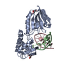
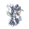
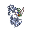
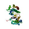
| Entry | Database: PDB / ID: 3saw | ||||||
|---|---|---|---|---|---|---|---|
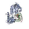
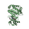
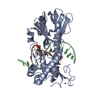
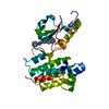
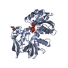
| Title | MUTM Slanted complex 8 with R112A mutation | ||||||
 Components Components |
| ||||||
 Keywords Keywords | HYDROLASE/DNA / DNA GLYCOSYLASE / DNA REPAIR / DAMAGE SEARCH / TRANSLOCATION / DISULFIDE CROSSLINKING / DNA DAMAGE / DNA-BINDING / GLYCOSIDASE / HYDROLASE / LYASE / METAL-BINDING / MULTIFUNCTIONAL ENZYME / ZINC-FINGER / HYDROLASE-DNA complex | ||||||
| Function / homology |  Function and homology information Function and homology informationDNA-formamidopyrimidine glycosylase / 8-oxo-7,8-dihydroguanine DNA N-glycosylase activity / class I DNA-(apurinic or apyrimidinic site) endonuclease activity / DNA-(apurinic or apyrimidinic site) lyase / base-excision repair / double-stranded DNA binding / damaged DNA binding / zinc ion binding Similarity search - Function | ||||||
| Biological species |   Geobacillus stearothermophilus (bacteria) Geobacillus stearothermophilus (bacteria) | ||||||
| Method |  X-RAY DIFFRACTION / SYNCHROTRON / MOLECULAR REPLACEMENT / Resolution: 2.35 Å X-RAY DIFFRACTION / SYNCHROTRON / MOLECULAR REPLACEMENT / Resolution: 2.35 Å | ||||||
 Authors Authors | Qi, Y. / Verdine, G.L. | ||||||
 Citation Citation | Journal: Proc.Natl.Acad.Sci.USA / Year: 2012 Title: Strandwise translocation of a DNA glycosylase on undamaged DNA. Authors: Qi, Y. / Nam, K. / Spong, M.C. / Banerjee, A. / Sung, R.J. / Zhang, M. / Karplus, M. / Verdine, G.L. | ||||||
| History |
|
- Structure visualization
| Structure viewer | Molecule: MolmilJmol/JSmol |
|---|
- Downloads & links
-Download
| PDBx/mmCIF format | 3saw.cif.gz | 140.3 KB | Display | PDBx/mmCIF format |
|---|---|---|---|---|
| PDB format | pdb3saw.ent.gz | 105.6 KB | Display | PDB format |
| PDBx/mmJSON format | 3saw.json.gz | Tree view | PDBx/mmJSON format | |
| Others |  Other downloads Other downloads |
-Validation report
| Arichive directory | https://data.pdbj.org/pub/pdb/validation_reports/sa/3sawftp://data.pdbj.org/pub/pdb/validation_reports/sa/3saw | HTTPS FTP |
|---|
-Related structure data
| Related structure data |  3sarC  3sasC  3satC  3sauC  3savC  3sbjC  2f5oS C: citing same article ( S: Starting model for refinement |
|---|---|
| Similar structure data |

-Links
 PDBj
PDBj


- Assembly
| Deposited unit | 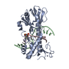
| ||||||||
|---|---|---|---|---|---|---|---|---|---|
| 1 |
| ||||||||
| Unit cell |
|
-Components
| #1: Protein | Mass: 30497.213 Da / Num. of mol.: 1 Source method: isolated from a genetically manipulated source Source: (gene. exp.) Geobacillus stearothermophilus (bacteria)Gene: MUTM / Plasmid: pET24B / Production host: References: UniProt: P84131, DNA-(apurinic or apyrimidinic site) lyase |
|---|---|
| #2: DNA chain | Mass: 4981.256 Da / Num. of mol.: 1 / Source method: obtained synthetically |
| #3: DNA chain | Mass: 4875.248 Da / Num. of mol.: 1 / Source method: obtained synthetically |
| #4: Chemical | ChemComp-ZN /   Mass: 65.409 Da / Num. of mol.: 1 / Source method: obtained synthetically / Formula: Zn Mass: 65.409 Da / Num. of mol.: 1 / Source method: obtained synthetically / Formula: Zn |
| #5: Water | ChemComp-HOH /  Mass: 18.015 Da / Num. of mol.: 98 / Source method: isolated from a natural source / Formula: H2O Mass: 18.015 Da / Num. of mol.: 98 / Source method: isolated from a natural source / Formula: H2O |
-Experimental details
-Experiment
| Experiment | Method: X-RAY DIFFRACTION / Number of used crystals: 1 |
|---|
- Sample preparation
| Crystal | Density Matthews: 2.81 Å3/Da / Density % sol: 56.15 % |
|---|---|
| Crystal grow | Temperature: 273 K / pH: 7 Details: PEG 8K, SODIUM CACODYLATE, GLYCEROL, PH 7.0, VAPOR DIFFUSION, HANGING DROP, TEMPERATURE 273K |
-Data collection
| Diffraction | Mean temperature: 100 K |
|---|---|
| Diffraction source | Source: SYNCHROTRON / Site: APS  / Beamline: 19-ID / Wavelength: 0.98 Å / Beamline: 19-ID / Wavelength: 0.98 Å |
| Detector | Type: ADSC QUANTUM 315 / Detector: CCD / Date: Aug 7, 2007 |
| Radiation | Protocol: SINGLE WAVELENGTH / Monochromatic (M) / Laue (L): M / Scattering type: x-ray |
| Radiation wavelength | Wavelength: 0.98 Å / Relative weight: 1 |
| Reflection | Resolution: 2.3→50 Å / Num. obs: 20503 / % possible obs: 98.2 % / Redundancy: 5 % / Rmerge(I) obs: 0.066 / Net I/σ(I): 11.9 |
| Reflection shell | Resolution: 2.3→2.38 Å / Redundancy: 5 % / Rmerge(I) obs: 0.417 / % possible all: 99.6 |
- Processing
| Software |
| |||||||||||||||||||||||||||||||||||||||||||||||||||||||||||||||||||||||||||
|---|---|---|---|---|---|---|---|---|---|---|---|---|---|---|---|---|---|---|---|---|---|---|---|---|---|---|---|---|---|---|---|---|---|---|---|---|---|---|---|---|---|---|---|---|---|---|---|---|---|---|---|---|---|---|---|---|---|---|---|---|---|---|---|---|---|---|---|---|---|---|---|---|---|---|---|---|
| Refinement | Method to determine structure: MOLECULAR REPLACEMENT Starting model: 2F5O Resolution: 2.35→26.3 Å / SU ML: 2.12 / σ(F): 0.19 / Stereochemistry target values: ML
| |||||||||||||||||||||||||||||||||||||||||||||||||||||||||||||||||||||||||||
| Solvent computation | Shrinkage radii: 0.9 Å / VDW probe radii: 1.11 Å / Solvent model: FLAT BULK SOLVENT MODEL / Bsol: 51.44 Å2 / ksol: 0.35 e/Å3 | |||||||||||||||||||||||||||||||||||||||||||||||||||||||||||||||||||||||||||
| Displacement parameters | Biso mean: 49.03 Å2
| |||||||||||||||||||||||||||||||||||||||||||||||||||||||||||||||||||||||||||
| Refinement step | Cycle: LAST / Resolution: 2.35→26.3 Å
| |||||||||||||||||||||||||||||||||||||||||||||||||||||||||||||||||||||||||||
| Refine LS restraints |
| |||||||||||||||||||||||||||||||||||||||||||||||||||||||||||||||||||||||||||
| LS refinement shell |
| |||||||||||||||||||||||||||||||||||||||||||||||||||||||||||||||||||||||||||
| Refinement TLS params. | Method: refined / Refine-ID: X-RAY DIFFRACTION
| |||||||||||||||||||||||||||||||||||||||||||||||||||||||||||||||||||||||||||
| Refinement TLS group |
|
