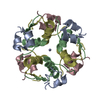
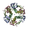
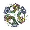
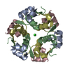
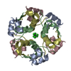
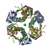
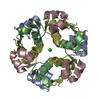
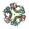
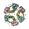
Acoustically mounted porcine insulin microcrystals yield an X-ray SAD structure
Components
(Insulin) x 2
Keywords
HORMONE
Function / homology
Function and homology information
positive regulation of lipoprotein lipase activity / Insulin processing / IRS activation / Signal attenuation / Insulin receptor signalling cascade / Signaling by Insulin receptor / Synthesis, secretion, and deacylation of Ghrelin / PI5P, PP2A and IER3 Regulate PI3K/AKT Signaling / Insulin receptor recycling / response to L-arginine ...positive regulation of lipoprotein lipase activity / Insulin processing / IRS activation / Signal attenuation / Insulin receptor signalling cascade / Signaling by Insulin receptor / Synthesis, secretion, and deacylation of Ghrelin / PI5P, PP2A and IER3 Regulate PI3K/AKT Signaling / Insulin receptor recycling / response to L-arginine / lactate biosynthetic process / positive regulation of glucose metabolic process / positive regulation of fatty acid biosynthetic process / glycoprotein biosynthetic process / lipoprotein biosynthetic process / COPI-mediated anterograde transport / negative regulation of glycogen catabolic process / : / negative regulation of fatty acid metabolic process / negative regulation of feeding behavior / lipid biosynthetic process / negative regulation of acute inflammatory response / positive regulation of respiratory burst / alpha-beta T cell activation / TORC1 signaling / negative regulation of protein secretion / positive regulation of dendritic spine maintenance / negative regulation of gluconeogenesis / fatty acid homeostasis / positive regulation of glycogen biosynthetic process / positive regulation of insulin receptor signaling pathway / negative regulation of respiratory burst involved in inflammatory response / negative regulation of lipid catabolic process / negative regulation of ubiquitin-dependent protein catabolic process / nitric oxide-cGMP-mediated signaling / regulation of protein localization to plasma membrane / negative regulation of reactive oxygen species biosynthetic process / insulin-like growth factor receptor binding / neuron projection maintenance / positive regulation of mitotic nuclear division / positive regulation of DNA replication / positive regulation of glycolytic process / positive regulation of cytokine production / acute-phase response / positive regulation of D-glucose import across plasma membrane / insulin receptor binding / positive regulation of protein secretion / positive regulation of translation / wound healing / hormone activity / positive regulation of neuron projection development / negative regulation of protein catabolic process / positive regulation of protein localization to nucleus / vasodilation / glucose metabolic process / insulin receptor signaling pathway / glucose homeostasis / protease binding / positive regulation of canonical NF-kappaB signal transduction / positive regulation of MAPK cascade / positive regulation of phosphatidylinositol 3-kinase/protein kinase B signal transduction / positive regulation of cell migration / G protein-coupled receptor signaling pathway / negative regulation of gene expression / positive regulation of cell population proliferation / : / identical protein binding Similarity search - Function
Insulin / Insulin family / Insulin-like / Insulin/IGF/Relaxin family / Insulin / insulin-like growth factor / relaxin family. / Insulin, conserved site / Insulin family signature. / Insulin-like superfamily Similarity search - Domain/homology
Mass: 18.015 Da / Num. of mol.: 46 / Source method: isolated from a natural source / Formula: H2O
Has protein modification
Y
-
Experimental details
-
Experiment
Experiment
Method: X-RAY DIFFRACTION / Number of used crystals: 9
-
Sample preparation
Crystal
Density Matthews: 1.88 Å3/Da / Density % sol: 34.42 %
Crystal grow
Method: small tubes / pH: 6 Details: Microcrystals are obtained by dissolving 0.025 g protein in 5 mL of crystallizing solution (200 mL 0.02 M HCl, 100 mL 0.20 M sodium citrate, 60 mL acetone, 20 mL water, 20 mL 0.12 M zinc ...Details: Microcrystals are obtained by dissolving 0.025 g protein in 5 mL of crystallizing solution (200 mL 0.02 M HCl, 100 mL 0.20 M sodium citrate, 60 mL acetone, 20 mL water, 20 mL 0.12 M zinc sulfate) at 315K. The solution is then rapidly quenched to 293K by immersing it in a cool water bath. Quenching speed determines the resulting crystal size. 20-micron crystals are obtained by quenching in a 293K water bath, 10-micron crystals by quenching in a 283K water bath, and 5-micron crystals by quenching in ice water. pH 6.0, SMALL TUBES
-
Data collection
Diffraction
ID
Mean temperature (K)
Crystal-ID
1
100
1
2
100
1
3
100
1
4
100
1
1,2,3,4
1
Diffraction source
Source
Site
Beamline
ID
Wavelength
SYNCHROTRON
NSLS
X12B
1
1.28
SYNCHROTRON
NSLS
X12C
2
1.28
SYNCHROTRON
NSLS
X25
3
1.28
SYNCHROTRON
APS
23-ID-D
4
0.9733
Detector
Type
ID
Detector
Date
ADSC QUANTUM 4
1
CCD
Apr 29, 2010
ADSC QUANTUM 210
2
CCD
Jul 30, 2010
ADSC QUANTUM 315
3
CCD
Feb 27, 2010
MARMOSAIC 300 mm CCD
4
CCD
Dec 19, 2009
Radiation
ID
Protocol
Monochromatic (M) / Laue (L)
Scattering type
Wavelength-ID
1
SINGLEWAVELENGTH
M
x-ray
1
2
SINGLEWAVELENGTH
M
x-ray
1
3
SINGLEWAVELENGTH
M
x-ray
1
4
SINGLEWAVELENGTH
M
x-ray
1
Radiation wavelength
ID
Wavelength (Å)
Relative weight
1
1.28
1
2
0.9733
1
Reflection
Resolution: 1.8→50 Å / Num. all: 7783 / Num. obs: 7770 / % possible obs: 99.8 % / Redundancy: 10.5 % / Rmerge(I) obs: 0.114 / Net I/σ(I): 62.2
-
Processing
Software
Name
Version
Classification
CBASS
datacollection
SHELXS
phasing
REFMAC
5.5.0109
refinement
HKL-2000
datareduction
HKL-2000
datascaling
Refinement
Method to determine structure: SAD Starting model: PDB ENTRY 4INS
Resolution: 1.8→40.85 Å / Cor.coef. Fo:Fc: 0.974 / Cor.coef. Fo:Fc free: 0.954 / SU B: 4.454 / SU ML: 0.131 / Cross valid method: THROUGHOUT / ESU R: 0.162 / ESU R Free: 0.143 / Stereochemistry target values: MAXIMUM LIKELIHOOD Details: HYDROGENS HAVE BEEN ADDED IN THE RIDING POSITIONS U VALUES : REFINED INDIVIDUALLY
Rfactor
Num. reflection
% reflection
Selection details
Rfree
0.22311
357
4.6 %
RANDOM
Rwork
0.18135
-
-
-
obs
0.18338
7413
99.83 %
-
all
-
7783
-
-
Solvent computation
Ion probe radii: 0.8 Å / Shrinkage radii: 0.8 Å / VDW probe radii: 1.4 Å / Solvent model: MASK
Displacement parameters
Biso mean: 37.509 Å2
Baniso -1
Baniso -2
Baniso -3
1-
0.18 Å2
0.09 Å2
0 Å2
2-
-
0.18 Å2
0 Å2
3-
-
-
-0.27 Å2
Refinement step
Cycle: LAST / Resolution: 1.8→40.85 Å
Protein
Nucleic acid
Ligand
Solvent
Total
Num. atoms
806
0
2
46
854
Refine LS restraints
Refine-ID
Type
Dev ideal
Dev ideal target
Number
X-RAY DIFFRACTION
r_bond_refined_d
0.023
0.022
889
X-RAY DIFFRACTION
r_bond_other_d
X-RAY DIFFRACTION
r_angle_refined_deg
2.784
1.963
1220
X-RAY DIFFRACTION
r_angle_other_deg
X-RAY DIFFRACTION
r_dihedral_angle_1_deg
6.773
5
115
X-RAY DIFFRACTION
r_dihedral_angle_2_deg
38.003
24.667
45
X-RAY DIFFRACTION
r_dihedral_angle_3_deg
17.954
15
145
X-RAY DIFFRACTION
r_dihedral_angle_4_deg
8.064
15
3
X-RAY DIFFRACTION
r_chiral_restr
0.134
0.2
134
X-RAY DIFFRACTION
r_gen_planes_refined
0.009
0.02
697
X-RAY DIFFRACTION
r_gen_planes_other
X-RAY DIFFRACTION
r_nbd_refined
X-RAY DIFFRACTION
r_nbd_other
X-RAY DIFFRACTION
r_nbtor_refined
X-RAY DIFFRACTION
r_nbtor_other
X-RAY DIFFRACTION
r_xyhbond_nbd_refined
X-RAY DIFFRACTION
r_xyhbond_nbd_other
X-RAY DIFFRACTION
r_metal_ion_refined
X-RAY DIFFRACTION
r_metal_ion_other
X-RAY DIFFRACTION
r_symmetry_vdw_refined
X-RAY DIFFRACTION
r_symmetry_vdw_other
X-RAY DIFFRACTION
r_symmetry_hbond_refined
X-RAY DIFFRACTION
r_symmetry_hbond_other
X-RAY DIFFRACTION
r_symmetry_metal_ion_refined
X-RAY DIFFRACTION
r_symmetry_metal_ion_other
X-RAY DIFFRACTION
r_mcbond_it
1.37
1.5
534
X-RAY DIFFRACTION
r_mcbond_other
X-RAY DIFFRACTION
r_mcangle_it
2.626
2
864
X-RAY DIFFRACTION
r_scbond_it
3.423
3
355
X-RAY DIFFRACTION
r_scangle_it
5.716
4.5
348
X-RAY DIFFRACTION
r_rigid_bond_restr
X-RAY DIFFRACTION
r_sphericity_free
X-RAY DIFFRACTION
r_sphericity_bonded
LS refinement shell
Resolution: 1.8→1.847 Å / Total num. of bins used: 20
Rfactor
Num. reflection
% reflection
Rfree
0.435
24
-
Rwork
0.314
511
-
obs
-
-
97.81 %
+
About Yorodumi
-
News
-
Feb 9, 2022. New format data for meta-information of EMDB entries
New format data for meta-information of EMDB entries
Version 3 of the EMDB header file is now the official format.
The previous official version 1.9 will be removed from the archive.
In the structure databanks used in Yorodumi, some data are registered as the other names, "COVID-19 virus" and "2019-nCoV". Here are the details of the virus and the list of structure data.
Jan 31, 2019. EMDB accession codes are about to change! (news from PDBe EMDB page)
EMDB accession codes are about to change! (news from PDBe EMDB page)
The allocation of 4 digits for EMDB accession codes will soon come to an end. Whilst these codes will remain in use, new EMDB accession codes will include an additional digit and will expand incrementally as the available range of codes is exhausted. The current 4-digit format prefixed with “EMD-” (i.e. EMD-XXXX) will advance to a 5-digit format (i.e. EMD-XXXXX), and so on. It is currently estimated that the 4-digit codes will be depleted around Spring 2019, at which point the 5-digit format will come into force.
The EM Navigator/Yorodumi systems omit the EMD- prefix.
Related info.:Q: What is EMD? / ID/Accession-code notation in Yorodumi/EM Navigator
Yorodumi is a browser for structure data from EMDB, PDB, SASBDB, etc.
This page is also the successor to EM Navigator detail page, and also detail information page/front-end page for Omokage search.
The word "yorodu" (or yorozu) is an old Japanese word meaning "ten thousand". "mi" (miru) is to see.
Related info.:EMDB / PDB / SASBDB / Comparison of 3 databanks / Yorodumi Search / Aug 31, 2016. New EM Navigator & Yorodumi / Yorodumi Papers / Jmol/JSmol / Function and homology information / Changes in new EM Navigator and Yorodumi
 Movie
Movie Controller

 Yorodumi
 Open data
 Basic information
 Components
 Keywords
 Function and homology information
 X-RAY DIFFRACTION /
  Authors
 Citation
 Structure visualization
 Downloads & links
 Other downloads
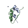

 PDBj
PDBj


 Assembly
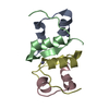


 Mass: 65.409 Da / Num. of mol.: 2 / Source method: obtained synthetically / Formula: Zn
 Mass: 18.015 Da / Num. of mol.: 46 / Source method: isolated from a natural source / Formula: H2O
 Sample preparation
 Processing