 Movie
Movie Controller

[English] 日本語
 Yorodumi
- PDB-3rqa: The Crystal Structure of a Pathogenic Protein from the Xanthomona... -
+ Open data

- Basic information
| Entry | Database: PDB / ID: 3rqa | ||||||
|---|---|---|---|---|---|---|---|
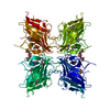
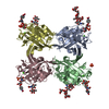
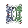
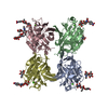
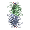
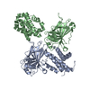
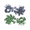
| Title | The Crystal Structure of a Pathogenic Protein from the Xanthomonas campestris Reveals a New Tetrameric PilZ Domain Self-Assembled via a Unusual Helical Bundle | ||||||
 Components Components | Putative uncharacterized protein | ||||||
 Keywords Keywords | UNKNOWN FUNCTION / PilZ domain / tetrameric parallel coiled-coil / four helix bundle | ||||||
| Function / homology | Cyclic di-GMP receptor, atypical PilZ domain / Atypical PilZ domain, cyclic di-GMP receptor / ACETATE ION / Cyclic di-GMP receptor atypical PilZ domain-containing protein Function and homology information Function and homology information | ||||||
| Biological species |  Xanthomonas campestris pv. campestris (bacteria) Xanthomonas campestris pv. campestris (bacteria) | ||||||
| Method |  X-RAY DIFFRACTION / SYNCHROTRON / MAD / Resolution: 2.1 Å X-RAY DIFFRACTION / SYNCHROTRON / MAD / Resolution: 2.1 Å | ||||||
 Authors Authors | Li, T.-N. / Chin, K.-H. / Chou, S.-H. | ||||||
 Citation Citation | Journal: Plos One / Year: 2011 Title: A novel tetrameric PilZ domain structure from xanthomonads Authors: Li, T.-N. / Chin, K.-H. / Fung, K.-M. / Yang, M.-T. / Wang, A.H.-J. / Chou, S.-H. | ||||||
| History |
|
- Structure visualization
| Structure viewer | Molecule: MolmilJmol/JSmol |
|---|
- Downloads & links
-Download
| PDBx/mmCIF format | 3rqa.cif.gz | 150.4 KB | Display | PDBx/mmCIF format |
|---|---|---|---|---|
| PDB format | pdb3rqa.ent.gz | 118.6 KB | Display | PDB format |
| PDBx/mmJSON format | 3rqa.json.gz | Tree view | PDBx/mmJSON format | |
| Others |  Other downloads Other downloads |
-Validation report
| Arichive directory | https://data.pdbj.org/pub/pdb/validation_reports/rq/3rqaftp://data.pdbj.org/pub/pdb/validation_reports/rq/3rqa | HTTPS FTP |
|---|
-Related structure data
| Similar structure data |
|---|

-Links
 PDBj
PDBj- Assembly
| Deposited unit | 
| ||||||||
|---|---|---|---|---|---|---|---|---|---|
| 1 |
| ||||||||
| Unit cell |
|
-Components
| #1: Protein | Mass: 20991.799 Da / Num. of mol.: 4 Source method: isolated from a genetically manipulated source Source: (gene. exp.) Xanthomonas campestris pv. campestris (bacteria)Strain: B100 / Gene: xcc-b100_2234 / Production host: #2: Chemical | ChemComp-ACT /   Mass: 59.044 Da / Num. of mol.: 4 / Source method: obtained synthetically / Formula: C2H3O2 Mass: 59.044 Da / Num. of mol.: 4 / Source method: obtained synthetically / Formula: C2H3O2#3: Water | ChemComp-HOH / |  Mass: 18.015 Da / Num. of mol.: 395 / Source method: isolated from a natural source / Formula: H2O Mass: 18.015 Da / Num. of mol.: 395 / Source method: isolated from a natural source / Formula: H2O |
|---|
-Experimental details
-Experiment
| Experiment | Method: X-RAY DIFFRACTION / Number of used crystals: 8 |
|---|
- Sample preparation
| Crystal | Density Matthews: 2.64 Å3/Da / Density % sol: 53.34 % Description: THE STRUCTURE FACTOR FILE CONTAINS FRIEDEL PAIRS |
|---|---|
| Crystal grow | Temperature: 298 K / Method: vapor diffusion, sitting drop / pH: 6.2 Details: 0.1M sodium/potassium phosphate, 0.2M NaCl, 10% (w/v) PEG 8000, pH 6.2, VAPOR DIFFUSION, SITTING DROP, temperature 298K |
-Data collection
| Diffraction | Mean temperature: 100 K | ||||||||||||
|---|---|---|---|---|---|---|---|---|---|---|---|---|---|
| Diffraction source | Source: SYNCHROTRON / Site: NSRRC  / Beamline: BL13B1 / Wavelength: 0.97910, 0.96388, 0.97884 / Beamline: BL13B1 / Wavelength: 0.97910, 0.96388, 0.97884 | ||||||||||||
| Detector | Type: ADSC QUANTUM 315r / Detector: CCD / Date: Jun 30, 2010 | ||||||||||||
| Radiation | Monochromator: Se / Protocol: MAD / Monochromatic (M) / Laue (L): M / Scattering type: x-ray | ||||||||||||
| Radiation wavelength |
| ||||||||||||
| Reflection | Resolution: 2.1→30 Å / Num. obs: 101046 / % possible obs: 98 % / Observed criterion σ(F): 2 / Observed criterion σ(I): 2 / Biso Wilson estimate: 18.8 Å2 | ||||||||||||
| Reflection shell | Resolution: 2.11→2.19 Å / % possible all: 98.4 |
- Processing
| Software |
| ||||||||||||||||||||||||||||||||||||||||||||||||||||||||||||||||||||||||||||||||
|---|---|---|---|---|---|---|---|---|---|---|---|---|---|---|---|---|---|---|---|---|---|---|---|---|---|---|---|---|---|---|---|---|---|---|---|---|---|---|---|---|---|---|---|---|---|---|---|---|---|---|---|---|---|---|---|---|---|---|---|---|---|---|---|---|---|---|---|---|---|---|---|---|---|---|---|---|---|---|---|---|---|
| Refinement | Method to determine structure: MAD / Resolution: 2.1→25.02 Å / Rfactor Rfree error: 0.004 / Data cutoff high absF: 500823.46 / Data cutoff low absF: 0 / Isotropic thermal model: RESTRAINED / Cross valid method: THROUGHOUT / σ(F): 0 Details: 1. BULK SOLVENT MODEL USED 2. SF FILE CONTAINS FRIEDEL PAIRS
| ||||||||||||||||||||||||||||||||||||||||||||||||||||||||||||||||||||||||||||||||
| Solvent computation | Solvent model: FLAT MODEL / Bsol: 47.0028 Å2 / ksol: 0.35 e/Å3 | ||||||||||||||||||||||||||||||||||||||||||||||||||||||||||||||||||||||||||||||||
| Displacement parameters | Biso mean: 44.3 Å2
| ||||||||||||||||||||||||||||||||||||||||||||||||||||||||||||||||||||||||||||||||
| Refine analyze |
| ||||||||||||||||||||||||||||||||||||||||||||||||||||||||||||||||||||||||||||||||
| Refinement step | Cycle: LAST / Resolution: 2.1→25.02 Å
| ||||||||||||||||||||||||||||||||||||||||||||||||||||||||||||||||||||||||||||||||
| Refine LS restraints |
| ||||||||||||||||||||||||||||||||||||||||||||||||||||||||||||||||||||||||||||||||
| LS refinement shell | Resolution: 2.1→2.23 Å / Rfactor Rfree error: 0.013 / Total num. of bins used: 6
| ||||||||||||||||||||||||||||||||||||||||||||||||||||||||||||||||||||||||||||||||
| Xplor file |
|
