 Movie
Movie Controller

[English] 日本語
 Yorodumi
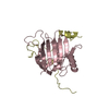
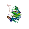
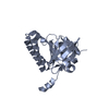
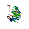
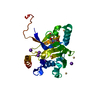
- PDB-3rq9: Structure of Tsi2, a Tse2-immunity protein from Pseudomonas aeruginosa -
+ Open data

- Basic information
| Entry | Database: PDB / ID: 3rq9 | ||||||
|---|---|---|---|---|---|---|---|
| Title | Structure of Tsi2, a Tse2-immunity protein from Pseudomonas aeruginosa | ||||||
 Components Components | Type VI secretion immunity protein | ||||||
 Keywords Keywords | TSE2-BINDING PROTEIN / immunity protein / type VI secretion / T6S / antitoxin | ||||||
| Function / homology | Helix Hairpins - #2500 / Tse2 immunity protein Tsi2 / : / : / T6SS, Immune protein Tsi2-like / Helix Hairpins / Orthogonal Bundle / Mainly Alpha / Immune protein Tsi2 Function and homology information Function and homology information | ||||||
| Biological species |   Pseudomonas aeruginosa (bacteria) Pseudomonas aeruginosa (bacteria) | ||||||
| Method |  X-RAY DIFFRACTION / SYNCHROTRON / MAD / Resolution: 1 Å X-RAY DIFFRACTION / SYNCHROTRON / MAD / Resolution: 1 Å | ||||||
 Authors Authors | Li, M. / Le Trong, I. / Stenkamp, R.E. / Mougous, J.D. | ||||||
 Citation Citation | Journal: Plos Pathog. / Year: 2012 Title: Structural Basis for Type VI Secretion Effector Recognition by a Cognate Immunity Protein. Authors: Li, M. / Le Trong, I. / Carl, M.A. / Larson, E.T. / Chou, S. / De Leon, J.A. / Dove, S.L. / Stenkamp, R.E. / Mougous, J.D. | ||||||
| History |
|
- Structure visualization
| Structure viewer | Molecule: MolmilJmol/JSmol |
|---|
- Downloads & links
-Download
| PDBx/mmCIF format | 3rq9.cif.gz | 84.6 KB | Display | PDBx/mmCIF format |
|---|---|---|---|---|
| PDB format | pdb3rq9.ent.gz | 66.3 KB | Display | PDB format |
| PDBx/mmJSON format | 3rq9.json.gz | Tree view | PDBx/mmJSON format | |
| Others |  Other downloads Other downloads |
-Validation report
| Arichive directory | https://data.pdbj.org/pub/pdb/validation_reports/rq/3rq9ftp://data.pdbj.org/pub/pdb/validation_reports/rq/3rq9 | HTTPS FTP |
|---|
-Related structure data
| Similar structure data |
|---|


-Links
 PDBj
PDBj- Assembly
| Deposited unit | 
| ||||||||
|---|---|---|---|---|---|---|---|---|---|
| 1 |
| ||||||||
| Unit cell |
|
-Components
| #1: Protein | Mass: 9595.624 Da / Num. of mol.: 2 Source method: isolated from a genetically manipulated source Source: (gene. exp.) Pseudomonas aeruginosa (bacteria) / Gene: PA2703 / Production host: #2: Water | ChemComp-HOH / |  Mass: 18.015 Da / Num. of mol.: 175 / Source method: isolated from a natural source / Formula: H2O Mass: 18.015 Da / Num. of mol.: 175 / Source method: isolated from a natural source / Formula: H2O |
|---|
-Experimental details
-Experiment
| Experiment | Method: X-RAY DIFFRACTION / Number of used crystals: 1 |
|---|
- Sample preparation
| Crystal | Density Matthews: 1.71 Å3/Da / Density % sol: 28.17 % |
|---|---|
| Crystal grow | Temperature: 298 K / Method: vapor diffusion, hanging drop / pH: 5 Details: 8% PEG 4000, 0.1M sodium acetate, pH 5.0, VAPOR DIFFUSION, HANGING DROP, temperature 298K |
-Data collection
| Diffraction | Mean temperature: 100 K | |||||||||
|---|---|---|---|---|---|---|---|---|---|---|
| Diffraction source | Source: SYNCHROTRON / Site: ALS  / Beamline: 8.2.2 / Wavelength: 0.9792, 0.9075 / Beamline: 8.2.2 / Wavelength: 0.9792, 0.9075 | |||||||||
| Detector | Type: ADSC QUANTUM 315 / Detector: CCD / Date: Nov 25, 2009 | |||||||||
| Radiation | Monochromator: double crystal, Si(111) / Protocol: MAD / Monochromatic (M) / Laue (L): M / Scattering type: x-ray | |||||||||
| Radiation wavelength |
| |||||||||
| Reflection | Resolution: 0.99→50 Å / Num. all: 60521 / Num. obs: 60521 / % possible obs: 100 % / Observed criterion σ(F): 0 / Observed criterion σ(I): 0 / Redundancy: 3.7 % / Rmerge(I) obs: 0.035 / Net I/σ(I): 31.2 | |||||||||
| Reflection shell | Resolution: 0.99→1.01 Å / Redundancy: 2.4 % / Rmerge(I) obs: 0.197 / Mean I/σ(I) obs: 3.7 / % possible all: 45 |
- Processing
| Software |
| ||||||||||||||||||||||||||||||||||||||||||||||||||||||||||||||||||||||||||||||||||||||||||||||||||||||||||||||||||||||||||||||||||||||||||||||||||||||||||||||||||||||||||
|---|---|---|---|---|---|---|---|---|---|---|---|---|---|---|---|---|---|---|---|---|---|---|---|---|---|---|---|---|---|---|---|---|---|---|---|---|---|---|---|---|---|---|---|---|---|---|---|---|---|---|---|---|---|---|---|---|---|---|---|---|---|---|---|---|---|---|---|---|---|---|---|---|---|---|---|---|---|---|---|---|---|---|---|---|---|---|---|---|---|---|---|---|---|---|---|---|---|---|---|---|---|---|---|---|---|---|---|---|---|---|---|---|---|---|---|---|---|---|---|---|---|---|---|---|---|---|---|---|---|---|---|---|---|---|---|---|---|---|---|---|---|---|---|---|---|---|---|---|---|---|---|---|---|---|---|---|---|---|---|---|---|---|---|---|---|---|---|---|---|---|---|
| Refinement | Method to determine structure: MAD / Resolution: 1→34.88 Å / Cor.coef. Fo:Fc: 0.973 / Cor.coef. Fo:Fc free: 0.963 / SU B: 0.701 / SU ML: 0.017 / Cross valid method: THROUGHOUT / ESU R: 0.028 / ESU R Free: 0.029 / Stereochemistry target values: MAXIMUM LIKELIHOOD / Details: HYDROGENS HAVE BEEN USED IF PRESENT IN THE INPUT
| ||||||||||||||||||||||||||||||||||||||||||||||||||||||||||||||||||||||||||||||||||||||||||||||||||||||||||||||||||||||||||||||||||||||||||||||||||||||||||||||||||||||||||
| Solvent computation | Ion probe radii: 0.8 Å / Shrinkage radii: 0.8 Å / VDW probe radii: 1.2 Å / Solvent model: BABINET MODEL WITH MASK | ||||||||||||||||||||||||||||||||||||||||||||||||||||||||||||||||||||||||||||||||||||||||||||||||||||||||||||||||||||||||||||||||||||||||||||||||||||||||||||||||||||||||||
| Displacement parameters | Biso mean: 10.798 Å2
| ||||||||||||||||||||||||||||||||||||||||||||||||||||||||||||||||||||||||||||||||||||||||||||||||||||||||||||||||||||||||||||||||||||||||||||||||||||||||||||||||||||||||||
| Refinement step | Cycle: LAST / Resolution: 1→34.88 Å
| ||||||||||||||||||||||||||||||||||||||||||||||||||||||||||||||||||||||||||||||||||||||||||||||||||||||||||||||||||||||||||||||||||||||||||||||||||||||||||||||||||||||||||
| Refine LS restraints |
| ||||||||||||||||||||||||||||||||||||||||||||||||||||||||||||||||||||||||||||||||||||||||||||||||||||||||||||||||||||||||||||||||||||||||||||||||||||||||||||||||||||||||||
| LS refinement shell | Resolution: 0.995→1.021 Å / Total num. of bins used: 20
|
