 Movie
Movie Controller


+ Open data

- Basic information
| Entry | Database: PDB / ID: 3vpv | ||||||
|---|---|---|---|---|---|---|---|
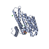
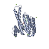
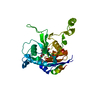
| Title | Crystal Structure of Pseudomonas aeruginosa Tsi2 | ||||||
 Components Components | Tse2 specific immunity protein 2 | ||||||
 Keywords Keywords | TOXIN INHIBITOR | ||||||
| Function / homology | Helix Hairpins - #2500 / Tse2 immunity protein Tsi2 / : / : / T6SS, Immune protein Tsi2-like / Helix Hairpins / Orthogonal Bundle / Mainly Alpha / Immune protein Tsi2 Function and homology information Function and homology information | ||||||
| Biological species |   Pseudomonas aeruginosa (bacteria) Pseudomonas aeruginosa (bacteria) | ||||||
| Method |  X-RAY DIFFRACTION / SYNCHROTRON / SAD / Resolution: 1.8 Å X-RAY DIFFRACTION / SYNCHROTRON / SAD / Resolution: 1.8 Å | ||||||
 Authors Authors | Wang, W. / Ding, J. / Wang, D.C. | ||||||
 Citation Citation | Journal: To be Published Title: Crystal Structure of Pseudomonas aeruginosa Tsi2 Authors: Wang, W. / Ding, J. / Wang, D.C. | ||||||
| History |
|
- Structure visualization
| Structure viewer | Molecule: MolmilJmol/JSmol |
|---|
- Downloads & links
-Download
| PDBx/mmCIF format | 3vpv.cif.gz | 48.1 KB | Display | PDBx/mmCIF format |
|---|---|---|---|---|
| PDB format | pdb3vpv.ent.gz | 34.2 KB | Display | PDB format |
| PDBx/mmJSON format | 3vpv.json.gz | Tree view | PDBx/mmJSON format | |
| Others |  Other downloads Other downloads |
-Validation report
| Arichive directory | https://data.pdbj.org/pub/pdb/validation_reports/vp/3vpvftp://data.pdbj.org/pub/pdb/validation_reports/vp/3vpv | HTTPS FTP |
|---|
-Related structure data
| Similar structure data |
|---|


-Links
 PDBj
PDBj- Assembly
| Deposited unit | 
| ||||||||
|---|---|---|---|---|---|---|---|---|---|
| 1 |
| ||||||||
| Unit cell |
|
-Components
| #1: Protein | Mass: 9689.414 Da / Num. of mol.: 2 Source method: isolated from a genetically manipulated source Source: (gene. exp.) Pseudomonas aeruginosa (bacteria) / Strain: ATCC 15692 / PAO1 / 1C / PRS 101 / LMG 12228 / Gene: PA2703 / Plasmid: pET22b / Production host: #2: Water | ChemComp-HOH / |  Mass: 18.015 Da / Num. of mol.: 218 / Source method: isolated from a natural source / Formula: H2O Mass: 18.015 Da / Num. of mol.: 218 / Source method: isolated from a natural source / Formula: H2OHas protein modification | Y | |
|---|
-Experimental details
-Experiment
| Experiment | Method: X-RAY DIFFRACTION / Number of used crystals: 1 |
|---|
- Sample preparation
| Crystal | Density Matthews: 1.82 Å3/Da / Density % sol: 32.27 % |
|---|---|
| Crystal grow | Temperature: 293 K / Method: vapor diffusion, sitting drop / pH: 5 Details: 10% PEG 3350, 0.1mM sodium acetate, pH 5.0, VAPOR DIFFUSION, SITTING DROP, temperature 293K |
-Data collection
| Diffraction | Mean temperature: 95 K |
|---|---|
| Diffraction source | Source: SYNCHROTRON / Site: BSRF  / Beamline: 3W1A / Wavelength: 0.9795 Å / Beamline: 3W1A / Wavelength: 0.9795 Å |
| Detector | Type: MAR CCD 165 mm / Detector: CCD / Date: Nov 14, 2011 |
| Radiation | Protocol: SINGLE WAVELENGTH / Monochromatic (M) / Laue (L): M / Scattering type: x-ray |
| Radiation wavelength | Wavelength: 0.9795 Å / Relative weight: 1 |
| Reflection | Resolution: 1.8→19.794 Å / Num. all: 12278 / Num. obs: 12200 / % possible obs: 99.4 % / Redundancy: 4 % / Biso Wilson estimate: 16.878 Å2 / Rmerge(I) obs: 0.022 / Rsym value: 0.022 / Net I/σ(I): 36.5 |
| Reflection shell | Resolution: 1.8→1.9 Å / Redundancy: 4 % / Rmerge(I) obs: 0.047 / Mean I/σ(I) obs: 16 / Num. unique all: 1771 / Rsym value: 0.047 / % possible all: 95.7 |
- Processing
| Software |
| ||||||||||||||||||||||||||||||
|---|---|---|---|---|---|---|---|---|---|---|---|---|---|---|---|---|---|---|---|---|---|---|---|---|---|---|---|---|---|---|---|
| Refinement | Method to determine structure: SAD / Resolution: 1.8→19.794 Å / Occupancy max: 1 / Occupancy min: 1 / FOM work R set: 0.8631 / SU ML: 0.21 / σ(F): 0.08 / Phase error: 21.31 / Stereochemistry target values: ML
| ||||||||||||||||||||||||||||||
| Solvent computation | Shrinkage radii: 0.72 Å / VDW probe radii: 1 Å / Solvent model: FLAT BULK SOLVENT MODEL / Bsol: 71.089 Å2 / ksol: 0.406 e/Å3 | ||||||||||||||||||||||||||||||
| Displacement parameters | Biso max: 43.61 Å2 / Biso mean: 16.23 Å2 / Biso min: 3.41 Å2
| ||||||||||||||||||||||||||||||
| Refine analyze | Luzzati coordinate error obs: 0.21 Å | ||||||||||||||||||||||||||||||
| Refinement step | Cycle: LAST / Resolution: 1.8→19.794 Å
| ||||||||||||||||||||||||||||||
| Refine LS restraints |
| ||||||||||||||||||||||||||||||
| LS refinement shell | Refine-ID: X-RAY DIFFRACTION / Total num. of bins used: 4
|
