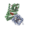
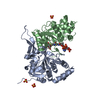
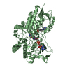
登録情報 データベース : PDB / ID : 3rl6タイトル Crystal structure of the archaeal asparagine synthetase A complexed with L-Asparagine and Adenosine monophosphate Archaeal asparagine synthetase A キーワード / / 機能・相同性 分子機能 ドメイン・相同性 構成要素
/ / / / / / / / / / / / / 生物種 Pyrococcus abyssi (古細菌)手法 / / / 解像度 : 2 Å データ登録者 Blaise, M. / Frechin, M. / Charron, C. / Roy, H. / Sauter, C. / Lorber, B. / Olieric, V. / Kern, D. ジャーナル : J.Mol.Biol. / 年 : 2011タイトル : Crystal Structure of the Archaeal Asparagine Synthetase: Interrelation with Aspartyl-tRNA and Asparaginyl-tRNA Synthetases.著者 : Blaise, M. / Frechin, M. / Olieric, V. / Charron, C. / Sauter, C. / Lorber, B. / Roy, H. / Kern, D. 履歴 登録 2011年4月19日 登録サイト / 処理サイト 改定 1.0 2011年8月17日 Provider / タイプ 改定 1.1 2011年9月21日 Group 改定 1.2 2024年2月28日 Group / Database references / Derived calculationsカテゴリ chem_comp_atom / chem_comp_bond ... chem_comp_atom / chem_comp_bond / database_2 / pdbx_struct_conn_angle / struct_conn / struct_site Item _database_2.pdbx_DOI / _database_2.pdbx_database_accession ... _database_2.pdbx_DOI / _database_2.pdbx_database_accession / _pdbx_struct_conn_angle.ptnr1_auth_comp_id / _pdbx_struct_conn_angle.ptnr1_auth_seq_id / _pdbx_struct_conn_angle.ptnr1_label_asym_id / _pdbx_struct_conn_angle.ptnr1_label_atom_id / _pdbx_struct_conn_angle.ptnr1_label_comp_id / _pdbx_struct_conn_angle.ptnr1_label_seq_id / _pdbx_struct_conn_angle.ptnr3_auth_comp_id / _pdbx_struct_conn_angle.ptnr3_auth_seq_id / _pdbx_struct_conn_angle.ptnr3_label_asym_id / _pdbx_struct_conn_angle.ptnr3_label_atom_id / _pdbx_struct_conn_angle.ptnr3_label_comp_id / _pdbx_struct_conn_angle.ptnr3_label_seq_id / _pdbx_struct_conn_angle.value / _struct_conn.pdbx_dist_value / _struct_conn.ptnr1_auth_comp_id / _struct_conn.ptnr1_auth_seq_id / _struct_conn.ptnr1_label_asym_id / _struct_conn.ptnr1_label_atom_id / _struct_conn.ptnr1_label_comp_id / _struct_conn.ptnr1_label_seq_id / _struct_conn.ptnr2_auth_comp_id / _struct_conn.ptnr2_auth_seq_id / _struct_conn.ptnr2_label_asym_id / _struct_conn.ptnr2_label_atom_id / _struct_conn.ptnr2_label_comp_id / _struct_site.pdbx_auth_asym_id / _struct_site.pdbx_auth_comp_id / _struct_site.pdbx_auth_seq_id
すべて表示 表示を減らす
 ムービー
ムービー コントローラー


 データを開く
 基本情報
基本情報 要素
要素 キーワード
キーワード 機能・相同性情報
 Pyrococcus abyssi (古細菌)
 X線回折 /
  データ登録者
 引用
引用 構造の表示
構造の表示 ダウンロードとリンク
 その他のダウンロード


 PDBj
PDBj


 集合体
集合体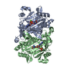

 タイプ: L-peptide linking / 分子量: 132.118 Da / 分子数: 2 / 由来タイプ: 合成 / 式: C4H8N2O3
 分子量: 347.221 Da / 分子数: 2 / 由来タイプ: 合成 / 式: C10H14N5O7P / コメント: AMP*YM
 分子量: 24.305 Da / 分子数: 1 / 由来タイプ: 合成 / 式: Mg
 分子量: 18.015 Da / 分子数: 334 / 由来タイプ: 天然 / 式: H2O
 試料調製
試料調製 / ビームライン: ID23-2 / 波長: 0.873 Å
 解析
解析