- PDB-3rk0: X-ray crystal Structure of the putative N-type ATP pyrophosphatas... -
+
Open data
ID or keywords:
Loading...
-
Basic information
Entry
Database: PDB / ID: 3rk0
Title
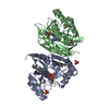
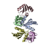
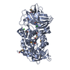
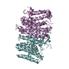
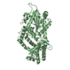
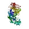
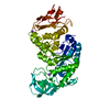
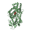
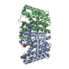
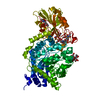
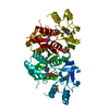
X-ray crystal Structure of the putative N-type ATP pyrophosphatase (PF0828) in complex with AMP from Pyrococcus furiosus, Northeast Structural Genomics Consortium Target PfR23
Components
N-type ATP pyrophosphatase superfamily
Keywords
HYDROLASE / Structural Genomics / PSI-Biology / Northeast Structural Genomics Consortium / NESG / alpha-beta protein that binds ATP / possibly n-type ATP pyrophosphatase / AMP and ATP binding
Function / homology
Function and homology information
diphthine-ammonia ligase activity / protein histidyl modification to diphthamide / ATP binding Similarity search - Function
Mass: 18.015 Da / Num. of mol.: 55 / Source method: isolated from a natural source / Formula: H2O
Has protein modification
Y
-
Experimental details
-
Experiment
Experiment
Method: X-RAY DIFFRACTION / Number of used crystals: 1
-
Sample preparation
Crystal
Density Matthews: 2.49 Å3/Da / Density % sol: 50.51 %
Crystal grow
Temperature: 291 K / Method: micro batch under oil / pH: 7.5 Details: Protein solution: 10 mM Tris (pH 7.5), 100 mM sodium chloride, 5 mM DTT, 0.02% NaN3, and 10mM dephospho-coenzyme A. Reservoir solution: 20% (w/v) PEG3350 and 200mM magnesium formate., micro ...Details: Protein solution: 10 mM Tris (pH 7.5), 100 mM sodium chloride, 5 mM DTT, 0.02% NaN3, and 10mM dephospho-coenzyme A. Reservoir solution: 20% (w/v) PEG3350 and 200mM magnesium formate., micro batch under oil, temperature 291K
In the structure databanks used in Yorodumi, some data are registered as the other names, "COVID-19 virus" and "2019-nCoV". Here are the details of the virus and the list of structure data.
Jan 31, 2019. EMDB accession codes are about to change! (news from PDBe EMDB page)
EMDB accession codes are about to change! (news from PDBe EMDB page)
The allocation of 4 digits for EMDB accession codes will soon come to an end. Whilst these codes will remain in use, new EMDB accession codes will include an additional digit and will expand incrementally as the available range of codes is exhausted. The current 4-digit format prefixed with “EMD-” (i.e. EMD-XXXX) will advance to a 5-digit format (i.e. EMD-XXXXX), and so on. It is currently estimated that the 4-digit codes will be depleted around Spring 2019, at which point the 5-digit format will come into force.
The EM Navigator/Yorodumi systems omit the EMD- prefix.
Related info.:Q: What is EMD? / ID/Accession-code notation in Yorodumi/EM Navigator
Yorodumi is a browser for structure data from EMDB, PDB, SASBDB, etc.
This page is also the successor to EM Navigator detail page, and also detail information page/front-end page for Omokage search.
The word "yorodu" (or yorozu) is an old Japanese word meaning "ten thousand". "mi" (miru) is to see.
Related info.:EMDB / PDB / SASBDB / Comparison of 3 databanks / Yorodumi Search / Aug 31, 2016. New EM Navigator & Yorodumi / Yorodumi Papers / Jmol/JSmol / Function and homology information / Changes in new EM Navigator and Yorodumi
 Movie
Movie Controller

 Yorodumi
 Open data
 Basic information
 Components
 Keywords
 Function and homology information
 Pyrococcus furiosus (archaea)
 X-RAY DIFFRACTION /
  Authors
 Citation
 Structure visualization
 Downloads & links
 Other downloads
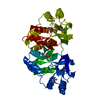

 PDBj
PDBj


 Assembly


 Mass: 347.221 Da / Num. of mol.: 1 / Source method: obtained synthetically / Formula: C10H14N5O7P / Comment: AMP*YM
 Mass: 18.015 Da / Num. of mol.: 55 / Source method: isolated from a natural source / Formula: H2O
 Sample preparation
 / Beamline: X4C / Wavelength: 0.97915 Å
 Processing