 Movie
Movie Controller

[English] 日本語
 Yorodumi
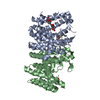
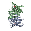
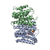
- PDB-5xn6: Heterodimer crystal structure of geranylgeranyl diphosphate synth... -
+ Open data

- Basic information
| Entry | Database: PDB / ID: 5xn6 | ||||||
|---|---|---|---|---|---|---|---|
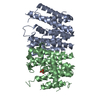
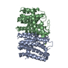
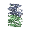
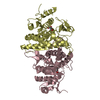
| Title | Heterodimer crystal structure of geranylgeranyl diphosphate synthases 1 with GGPPS Recruiting Protein(OsGRP) from Oryza sativa | ||||||
 Components Components |
| ||||||
 Keywords Keywords | TRANSFERASE / heterodimer / small-subunit / OsGGPPS1 / OsGRP | ||||||
| Function / homology |  Function and homology information Function and homology informationprenyltransferase activity / geranylgeranyl diphosphate synthase activity / isoprenoid biosynthetic process / metal ion binding / cytoplasm Similarity search - Function | ||||||
| Biological species |  | ||||||
| Method |  X-RAY DIFFRACTION / SYNCHROTRON / MOLECULAR REPLACEMENT / Resolution: 3.598 Å X-RAY DIFFRACTION / SYNCHROTRON / MOLECULAR REPLACEMENT / Resolution: 3.598 Å | ||||||
 Authors Authors | Wang, C. / Zhou, F. / Lu, S. / Zhang, P. | ||||||
| Funding support |  China, 1items China, 1items
| ||||||
 Citation Citation | Journal: Proc. Natl. Acad. Sci. U.S.A. / Year: 2017 Title: A recruiting protein of geranylgeranyl diphosphate synthase controls metabolic flux toward chlorophyll biosynthesis in rice Authors: Zhou, F. / Wang, C.Y. / Gutensohn, M. / Jiang, L. / Zhang, P. / Zhang, D. / Dudareva, N. / Lu, S. | ||||||
| History |
|
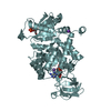
- Structure visualization
| Structure viewer | Molecule: MolmilJmol/JSmol |
|---|
- Downloads & links
-Download
| PDBx/mmCIF format | 5xn6.cif.gz | 594.7 KB | Display | PDBx/mmCIF format |
|---|---|---|---|---|
| PDB format | pdb5xn6.ent.gz | 501.3 KB | Display | PDB format |
| PDBx/mmJSON format | 5xn6.json.gz | Tree view | PDBx/mmJSON format | |
| Others |  Other downloads Other downloads |
-Validation report
| Arichive directory | https://data.pdbj.org/pub/pdb/validation_reports/xn/5xn6ftp://data.pdbj.org/pub/pdb/validation_reports/xn/5xn6 | HTTPS FTP |
|---|
-Related structure data
| Related structure data |  5xn5C 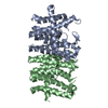 5e8hS S: Starting model for refinement C: citing same article ( |
|---|---|
| Similar structure data |
-Links
 PDBj
PDBj
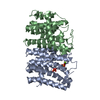
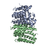
- Assembly
| Deposited unit | 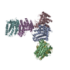
| ||||||||
|---|---|---|---|---|---|---|---|---|---|
| 1 | 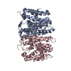
| ||||||||
| 2 | 
| ||||||||
| 3 | 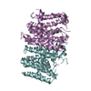
| ||||||||
| Unit cell |
|
-Components
| #1: Protein | Mass: 32865.441 Da / Num. of mol.: 3 / Fragment: UNP residues 62-366 Source method: isolated from a genetically manipulated source Source: (gene. exp.) Gene: Os07g0580900, OJ1301_C12.3, OSNPB_070580900, P0453G03.27 Production host:  #2: Protein | Mass: 32127.785 Da / Num. of mol.: 3 / Fragment: UNP residues 39-342 Source method: isolated from a genetically manipulated source Source: (gene. exp.) Gene: Os02g0668100, OJ1725_H08.13, OSNPB_020668100 Production host: References: UniProt: Q6ET88 |
|---|
-Experimental details
-Experiment
| Experiment | Method: X-RAY DIFFRACTION / Number of used crystals: 1 |
|---|
- Sample preparation
| Crystal | Density Matthews: 4.35 Å3/Da / Density % sol: 71.74 % |
|---|---|
| Crystal grow | Temperature: 293 K / Method: vapor diffusion, sitting drop / pH: 6.5 / Details: 16% PEG 10000, 0.1 M Bis-Tris, pH 6.5 |
-Data collection
| Diffraction | Mean temperature: 77 K |
|---|---|
| Diffraction source | Source: SYNCHROTRON / Site: SSRF / Beamline: BL17B1 / Wavelength: 0.9798 Å |
| Detector | Type: CS-PAD CXI-1 / Detector: PIXEL / Date: Sep 10, 2013 |
| Radiation | Protocol: SINGLE WAVELENGTH / Monochromatic (M) / Laue (L): M / Scattering type: x-ray |
| Radiation wavelength | Wavelength: 0.9798 Å / Relative weight: 1 |
| Reflection | Resolution: 3.59→37.8 Å / Num. obs: 36920 / % possible obs: 96.9 % / Redundancy: 1.9 % / Net I/σ(I): 8.26 |
- Processing
| Software |
| ||||||||||||||||||||||||||||||||||||||||||||||||||||||||||||||||||||||||||||||||||||||||||||||||||
|---|---|---|---|---|---|---|---|---|---|---|---|---|---|---|---|---|---|---|---|---|---|---|---|---|---|---|---|---|---|---|---|---|---|---|---|---|---|---|---|---|---|---|---|---|---|---|---|---|---|---|---|---|---|---|---|---|---|---|---|---|---|---|---|---|---|---|---|---|---|---|---|---|---|---|---|---|---|---|---|---|---|---|---|---|---|---|---|---|---|---|---|---|---|---|---|---|---|---|---|
| Refinement | Method to determine structure: MOLECULAR REPLACEMENT Starting model: 5E8H Resolution: 3.598→37.755 Å / SU ML: 0.54 / Cross valid method: FREE R-VALUE / σ(F): 1.96 / Phase error: 30.7 / Stereochemistry target values: ML
| ||||||||||||||||||||||||||||||||||||||||||||||||||||||||||||||||||||||||||||||||||||||||||||||||||
| Solvent computation | Shrinkage radii: 0.9 Å / VDW probe radii: 1.11 Å / Solvent model: FLAT BULK SOLVENT MODEL | ||||||||||||||||||||||||||||||||||||||||||||||||||||||||||||||||||||||||||||||||||||||||||||||||||
| Displacement parameters | Biso max: 150.48 Å2 / Biso mean: 90.3147 Å2 / Biso min: 30 Å2 | ||||||||||||||||||||||||||||||||||||||||||||||||||||||||||||||||||||||||||||||||||||||||||||||||||
| Refinement step | Cycle: final / Resolution: 3.598→37.755 Å
| ||||||||||||||||||||||||||||||||||||||||||||||||||||||||||||||||||||||||||||||||||||||||||||||||||
| Refine LS restraints |
| ||||||||||||||||||||||||||||||||||||||||||||||||||||||||||||||||||||||||||||||||||||||||||||||||||
| LS refinement shell | Refine-ID: X-RAY DIFFRACTION / Rfactor Rfree error: 0 / Total num. of bins used: 13
| ||||||||||||||||||||||||||||||||||||||||||||||||||||||||||||||||||||||||||||||||||||||||||||||||||
| Refinement TLS params. | Method: refined / Origin x: 17.4289 Å / Origin y: 91.8222 Å / Origin z: 84.0313 Å
| ||||||||||||||||||||||||||||||||||||||||||||||||||||||||||||||||||||||||||||||||||||||||||||||||||
| Refinement TLS group |
|
