 Movie
Movie Controller

[English] 日本語
 Yorodumi
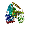
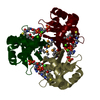
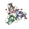
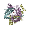
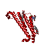
- PDB-3r3t: Crystal Structure of 30S Ribosomal Protein S from Bacillus anthracis -
+ Open data

- Basic information
| Entry | Database: PDB / ID: 3r3t | ||||||
|---|---|---|---|---|---|---|---|
| Title | Crystal Structure of 30S Ribosomal Protein S from Bacillus anthracis | ||||||
 Components Components | 30S ribosomal protein S6 | ||||||
 Keywords Keywords | RNA BINDING PROTEIN / Structural Genomics / Center for Structural Genomics of Infectious Diseases / CSGID / beta-barrel / cytosol | ||||||
| Function / homology |  Function and homology information Function and homology informationrRNA binding / ribosome / structural constituent of ribosome / translation Similarity search - Function | ||||||
| Biological species |  | ||||||
| Method |  X-RAY DIFFRACTION / SYNCHROTRON / SAD / Resolution: 2.302 Å X-RAY DIFFRACTION / SYNCHROTRON / SAD / Resolution: 2.302 Å | ||||||
 Authors Authors | Kim, Y. / Zhou, M. / Kwon, K. / Anderson, W.F. / Joachimiak, A. / Center for Structural Genomics of Infectious Diseases (CSGID) | ||||||
 Citation Citation | Journal: To be Published Title: Crystal Structure of 30S Ribosomal Protein S from Bacillus anthracis Authors: Kim, Y. / Zhou, M. / Kwon, K. / Anderson, W.F. / Joachimiak, A. / Center for Structural Genomics of Infectious Diseases (CSGID) | ||||||
| History |
|
- Structure visualization
| Structure viewer | Molecule: MolmilJmol/JSmol |
|---|
- Downloads & links
-Download
| PDBx/mmCIF format | 3r3t.cif.gz | 86.1 KB | Display | PDBx/mmCIF format |
|---|---|---|---|---|
| PDB format | pdb3r3t.ent.gz | 71.5 KB | Display | PDB format |
| PDBx/mmJSON format | 3r3t.json.gz | Tree view | PDBx/mmJSON format | |
| Others |  Other downloads Other downloads |
-Validation report
| Summary document | 3r3t_validation.pdf.gz | 459.7 KB | Display | wwPDB validaton report |
|---|---|---|---|---|
| Full document | 3r3t_full_validation.pdf.gz | 465.5 KB | Display | |
| Data in XML | 3r3t_validation.xml.gz | 10.3 KB | Display | |
| Data in CIF | 3r3t_validation.cif.gz | 12.9 KB | Display | |
| Arichive directory | https://data.pdbj.org/pub/pdb/validation_reports/r3/3r3tftp://data.pdbj.org/pub/pdb/validation_reports/r3/3r3t | HTTPS FTP |
-Related structure data
| Similar structure data | |
|---|---|
| Other databases |


-Links
 PDBj
PDBj- Assembly
| Deposited unit | 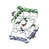
| |||||||||
|---|---|---|---|---|---|---|---|---|---|---|
| 1 | 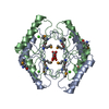
| |||||||||
| Unit cell |
| |||||||||
| Components on special symmetry positions |
|
-Components
| #1: Protein | Mass: 11697.859 Da / Num. of mol.: 2 Source method: isolated from a genetically manipulated source Source: (gene. exp.) #2: Chemical |   Mass: 40.078 Da / Num. of mol.: 2 / Source method: obtained synthetically / Formula: Ca Mass: 40.078 Da / Num. of mol.: 2 / Source method: obtained synthetically / Formula: Ca#3: Chemical | ChemComp-GOL / |   Mass: 92.094 Da / Num. of mol.: 1 / Source method: obtained synthetically / Formula: C3H8O3 Mass: 92.094 Da / Num. of mol.: 1 / Source method: obtained synthetically / Formula: C3H8O3#4: Chemical | ChemComp-SO4 / |   Mass: 96.063 Da / Num. of mol.: 1 / Source method: obtained synthetically / Formula: SO4 Mass: 96.063 Da / Num. of mol.: 1 / Source method: obtained synthetically / Formula: SO4#5: Water | ChemComp-HOH / |  Mass: 18.015 Da / Num. of mol.: 41 / Source method: isolated from a natural source / Formula: H2O Mass: 18.015 Da / Num. of mol.: 41 / Source method: isolated from a natural source / Formula: H2O |
|---|
-Experimental details
-Experiment
| Experiment | Method: X-RAY DIFFRACTION / Number of used crystals: 1 |
|---|
- Sample preparation
| Crystal | Density Matthews: 2.5 Å3/Da / Density % sol: 50.71 % |
|---|---|
| Crystal grow | Temperature: 289 K / Method: vapor diffusion, sitting drop / pH: 7.5 Details: 50 mM magnesium chloride, 0.1 M HEPES, 30 % v/v polyethylene glycol monomethyl ether 550, pH 7.5, VAPOR DIFFUSION, SITTING DROP, temperature 289K |
-Data collection
| Diffraction | Mean temperature: 100 K |
|---|---|
| Diffraction source | Source: SYNCHROTRON / Site: APS  / Beamline: 19-ID / Wavelength: 0.97942 Å / Beamline: 19-ID / Wavelength: 0.97942 Å |
| Detector | Type: ADSC QUANTUM 315r / Detector: CCD / Date: Oct 13, 2010 / Details: mirrors |
| Radiation | Monochromator: Si(111) / Protocol: SAD / Monochromatic (M) / Laue (L): M / Scattering type: x-ray |
| Radiation wavelength | Wavelength: 0.97942 Å / Relative weight: 1 |
| Reflection | Resolution: 2.3→50 Å / Num. all: 10636 / Num. obs: 10636 / % possible obs: 99.8 % / Observed criterion σ(F): 0 / Observed criterion σ(I): 0 / Redundancy: 12.9 % / Biso Wilson estimate: 51.15 Å2 / Rsym value: 0.105 / Net I/σ(I): 10.3 |
| Reflection shell | Resolution: 2.3→2.34 Å / Redundancy: 13.2 % / Mean I/σ(I) obs: 4.65 / Num. unique all: 511 / Rsym value: 0.621 / % possible all: 100 |
- Processing
| Software |
| |||||||||||||||||||||||||||||||||||||||||||||||||||||||||||||||||||||||||||
|---|---|---|---|---|---|---|---|---|---|---|---|---|---|---|---|---|---|---|---|---|---|---|---|---|---|---|---|---|---|---|---|---|---|---|---|---|---|---|---|---|---|---|---|---|---|---|---|---|---|---|---|---|---|---|---|---|---|---|---|---|---|---|---|---|---|---|---|---|---|---|---|---|---|---|---|---|
| Refinement | Method to determine structure: SAD / Resolution: 2.302→45.609 Å / Isotropic thermal model: mixed / Cross valid method: throughput / σ(F): 0 / Phase error: 25.5 / Stereochemistry target values: TWIN_LSQ_F
| |||||||||||||||||||||||||||||||||||||||||||||||||||||||||||||||||||||||||||
| Solvent computation | Shrinkage radii: 1.06 Å / VDW probe radii: 1.3 Å / Solvent model: FLAT BULK SOLVENT MODEL / Bsol: 58.712 Å2 / ksol: 0.359 e/Å3 | |||||||||||||||||||||||||||||||||||||||||||||||||||||||||||||||||||||||||||
| Displacement parameters | Biso mean: 51.2 Å2
| |||||||||||||||||||||||||||||||||||||||||||||||||||||||||||||||||||||||||||
| Refinement step | Cycle: LAST / Resolution: 2.302→45.609 Å
| |||||||||||||||||||||||||||||||||||||||||||||||||||||||||||||||||||||||||||
| Refine LS restraints |
| |||||||||||||||||||||||||||||||||||||||||||||||||||||||||||||||||||||||||||
| Refinement TLS params. | Method: refined / Refine-ID: X-RAY DIFFRACTION
| |||||||||||||||||||||||||||||||||||||||||||||||||||||||||||||||||||||||||||
| Refinement TLS group |
|
