| ソフトウェア | | 名称 | バージョン | 分類 |
|---|
| SBC-Collect | | データ収集 | | SHELXD | | 位相決定 | | MLPHARE | | 位相決定 | | 直接法 | | モデル構築 | | ARP | | モデル構築 | | WARP | | モデル構築 | | HKL-3000 | | 位相決定 | | PHENIX | (phenix.refine: 1.6.4_486)| 精密化 | | HKL-3000 | | データ削減 | | HKL-3000 | | データスケーリング | | 直接法 | | 位相決定 | |
|
|---|
| 精密化 | 構造決定の手法:  単波長異常分散 / 解像度: 1.383→37.076 Å / SU ML: 0.15 / σ(F): 0 / 位相誤差: 17.8 / 立体化学のターゲット値: ML 単波長異常分散 / 解像度: 1.383→37.076 Å / SU ML: 0.15 / σ(F): 0 / 位相誤差: 17.8 / 立体化学のターゲット値: ML
| Rfactor | 反射数 | %反射 | Selection details |
|---|
| Rfree | 0.1851 | 2141 | 5.05 % | random |
|---|
| Rwork | 0.1634 | - | - | - |
|---|
| obs | 0.1645 | 42410 | 95.9 % | - |
|---|
| all | - | 42410 | - | - |
|---|
|
|---|
| 溶媒の処理 | 減衰半径: 0.61 Å / VDWプローブ半径: 0.9 Å / 溶媒モデル: FLAT BULK SOLVENT MODEL / Bsol: 60.231 Å2 / ksol: 0.375 e/Å3 |
|---|
| 原子変位パラメータ | | Baniso -1 | Baniso -2 | Baniso -3 |
|---|
| 1- | 3.4919 Å2 | 0 Å2 | -0 Å2 |
|---|
| 2- | - | 0.3151 Å2 | 0 Å2 |
|---|
| 3- | - | - | -3.807 Å2 |
|---|
|
|---|
| 精密化ステップ | サイクル: LAST / 解像度: 1.383→37.076 Å
| タンパク質 | 核酸 | リガンド | 溶媒 | 全体 |
|---|
| 原子数 | 1897 | 0 | 24 | 299 | 2220 |
|---|
|
|---|
| 拘束条件 | | Refine-ID | タイプ | Dev ideal | 数 |
|---|
| X-RAY DIFFRACTION | f_bond_d| 0.007 | 2043 | | X-RAY DIFFRACTION | f_angle_d| 1.178 | 2815 | | X-RAY DIFFRACTION | f_dihedral_angle_d| 11.945 | 762 | | X-RAY DIFFRACTION | f_chiral_restr| 0.073 | 317 | | X-RAY DIFFRACTION | f_plane_restr| 0.009 | 380 | | | | | |
|
|---|
| LS精密化 シェル | | 解像度 (Å) | Rfactor Rfree | Num. reflection Rfree | Rfactor Rwork | Num. reflection Rwork | Refine-ID | % reflection obs (%) |
|---|
| 1.383-1.4324 | 0.2782 | 177 | 0.257 | 3433 | X-RAY DIFFRACTION | 83 | | 1.4324-1.4898 | 0.2695 | 192 | 0.2226 | 3743 | X-RAY DIFFRACTION | 90 | | 1.4898-1.5576 | 0.2369 | 209 | 0.1904 | 3899 | X-RAY DIFFRACTION | 94 | | 1.5576-1.6397 | 0.1879 | 220 | 0.1637 | 3961 | X-RAY DIFFRACTION | 96 | | 1.6397-1.7424 | 0.2024 | 221 | 0.1614 | 4063 | X-RAY DIFFRACTION | 98 | | 1.7424-1.877 | 0.1744 | 215 | 0.1622 | 4141 | X-RAY DIFFRACTION | 99 | | 1.877-2.0658 | 0.1799 | 240 | 0.1565 | 4138 | X-RAY DIFFRACTION | 100 | | 2.0658-2.3647 | 0.1664 | 226 | 0.1596 | 4209 | X-RAY DIFFRACTION | 100 | | 2.3647-2.9791 | 0.1901 | 213 | 0.1688 | 4252 | X-RAY DIFFRACTION | 100 | | 2.9791-37.0892 | 0.1706 | 228 | 0.1495 | 4430 | X-RAY DIFFRACTION | 100 |
|
|---|
| 精密化 TLS | 手法: refined / Origin x: 47.8165 Å / Origin y: 10.1777 Å / Origin z: 5.0956 Å
| 11 | 12 | 13 | 21 | 22 | 23 | 31 | 32 | 33 |
|---|
| T | 0.1088 Å2 | -0.0034 Å2 | 0.0003 Å2 | - | 0.1148 Å2 | 0.0009 Å2 | - | - | 0.1128 Å2 |
|---|
| L | 0.1188 °2 | 0.0398 °2 | -0.0017 °2 | - | 0.2175 °2 | 0.0767 °2 | - | - | 0.0361 °2 |
|---|
| S | 0.0167 Å ° | -0.0054 Å ° | 0.0086 Å ° | 0.0023 Å ° | -0.0082 Å ° | 0.0049 Å ° | -0.0002 Å ° | 0.0024 Å ° | -0.0043 Å ° |
|---|
|
|---|
| 精密化 TLSグループ | Selection details: chain A |
|---|
 ムービー
ムービー コントローラー


 データを開く
 基本情報
基本情報 要素
要素 キーワード
キーワード 機能・相同性情報
 Sphaerobacter thermophilus (バクテリア)
 データ登録者
 引用
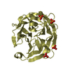
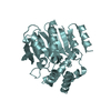
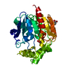
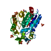
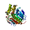
引用 構造の表示
構造の表示 ダウンロードとリンク
 その他のダウンロード
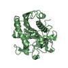


 PDBj
PDBj


 集合体
集合体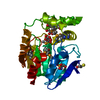


 分子量: 35.453 Da / 分子数: 1 / 由来タイプ: 合成 / 式: Cl
 分子量: 92.094 Da / 分子数: 1 / 由来タイプ: 合成 / 式: C3H8O3
 分子量: 78.133 Da / 分子数: 2 / 由来タイプ: 合成 / 式: C2H6OS
 分子量: 134.087 Da / 分子数: 1 / 由来タイプ: 合成 / 式: C4H6O5
 試料調製
試料調製 / ビームライン: 19-ID / 波長: 0.97918 Å
 解析
解析