 Movie
Movie Controller

[English] 日本語
 Yorodumi
- PDB-6ra2: Structural basis for recognition and ring-cleavage of the Pseudom... -
+ Open data

- Basic information
| Entry | Database: PDB / ID: 6ra2 | |||||||||
|---|---|---|---|---|---|---|---|---|---|---|
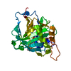
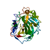
| Title | Structural basis for recognition and ring-cleavage of the Pseudomonas quinolone signal (PQS) by AqDC | |||||||||
 Components Components | Putative dioxygenase (1H-3-hydroxy-4-oxoquinaldine 2,4-dioxygenase) | |||||||||
 Keywords Keywords | HYDROLASE / dioxygenase / alpha/beta hydrolase fold / catalytic triad / quorum sensing / Pseudomonas quinolone signal / Pseudomonas aeruginosa | |||||||||
| Function / homology |  Function and homology information Function and homology informationOxidoreductases; Acting on single donors with incorporation of molecular oxygen (oxygenases); With incorporation of two atoms of oxygen / dioxygenase activity Similarity search - Function | |||||||||
| Biological species |  Mycobacteroides abscessus (bacteria) Mycobacteroides abscessus (bacteria) | |||||||||
| Method |  X-RAY DIFFRACTION / SYNCHROTRON / MOLECULAR REPLACEMENT / Resolution: 2.3 Å X-RAY DIFFRACTION / SYNCHROTRON / MOLECULAR REPLACEMENT / Resolution: 2.3 Å | |||||||||
 Authors Authors | Wullich, S. / Kobus, S. / Smits, S.H. / Fetzner, S. | |||||||||
 Citation Citation | Journal: J.Struct.Biol. / Year: 2019 Title: Structural basis for recognition and ring-cleavage of the Pseudomonas quinolone signal (PQS) by AqdC, a mycobacterial dioxygenase of the alpha / beta-hydrolase fold family. Authors: Wullich, S.C. / Kobus, S. / Wienhold, M. / Hennecke, U. / Smits, S.H.J. / Fetzner, S. | |||||||||
| History |
|
- Structure visualization
| Structure viewer | Molecule: MolmilJmol/JSmol |
|---|
- Downloads & links
-Download
| PDBx/mmCIF format | 6ra2.cif.gz | 174 KB | Display | PDBx/mmCIF format |
|---|---|---|---|---|
| PDB format | pdb6ra2.ent.gz | 137.9 KB | Display | PDB format |
| PDBx/mmJSON format | 6ra2.json.gz | Tree view | PDBx/mmJSON format | |
| Others |  Other downloads Other downloads |
-Validation report
| Arichive directory | https://data.pdbj.org/pub/pdb/validation_reports/ra/6ra2ftp://data.pdbj.org/pub/pdb/validation_reports/ra/6ra2 | HTTPS FTP |
|---|
-Related structure data
| Related structure data |  6ra3C  6rb3C  2wj3S S: Starting model for refinement C: citing same article ( |
|---|---|
| Similar structure data |


-Links
 PDBj
PDBj


- Assembly
| Deposited unit | 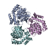
| ||||||||
|---|---|---|---|---|---|---|---|---|---|
| 1 | 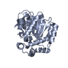
| ||||||||
| 2 | 
| ||||||||
| 3 | 
| ||||||||
| Unit cell |
| ||||||||
| Components on special symmetry positions |
|
-Components
| #1: Protein | Mass: 29797.703 Da / Num. of mol.: 3 Source method: isolated from a genetically manipulated source Source: (gene. exp.) Mycobacteroides abscessus (bacteria) / Gene: MAB_0303 / Production host: #2: Water | ChemComp-HOH / |  Mass: 18.015 Da / Num. of mol.: 563 / Source method: isolated from a natural source / Formula: H2O Mass: 18.015 Da / Num. of mol.: 563 / Source method: isolated from a natural source / Formula: H2O |
|---|
-Experimental details
-Experiment
| Experiment | Method: X-RAY DIFFRACTION / Number of used crystals: 1 |
|---|
- Sample preparation
| Crystal | Density Matthews: 2.49 Å3/Da / Density % sol: 50.57 % |
|---|---|
| Crystal grow | Temperature: 298 K / Method: vapor diffusion, hanging drop / pH: 6.5 Details: 0.10 to 0.15 M magnesium chloride, 0.1 M sodium chloride, 0.1 M MES pH 6.5 and 30 to 40 % PEG 400 at 12C. PH range: 6.2-6.8 |
-Data collection
| Diffraction | Mean temperature: 100 K / Serial crystal experiment: N |
|---|---|
| Diffraction source | Source: SYNCHROTRON / Site: ESRF  / Beamline: ID23-2 / Wavelength: 0.97625 Å / Beamline: ID23-2 / Wavelength: 0.97625 Å |
| Detector | Type: DECTRIS PILATUS3 R CdTe 300K / Detector: PIXEL / Date: Jul 15, 2018 |
| Radiation | Protocol: SINGLE WAVELENGTH / Monochromatic (M) / Laue (L): M / Scattering type: x-ray |
| Radiation wavelength | Wavelength: 0.97625 Å / Relative weight: 1 |
| Reflection | Resolution: 2.3→46.91 Å / Num. obs: 40361 / % possible obs: 99.3 % / Redundancy: 7.4 % / CC1/2: 0.998 / Rmerge(I) obs: 0.06186 / Net I/σ(I): 29.82 |
| Reflection shell | Resolution: 2.3→2.38 Å / Rmerge(I) obs: 0.1996 / Num. unique obs: 3990 / CC1/2: 0.984 |
- Processing
| Software |
| ||||||||||||||||||||||||||||||||||||||||||||||||||||||||||||||||||||||||||||||||||||||||||||||||||||||||||||||||
|---|---|---|---|---|---|---|---|---|---|---|---|---|---|---|---|---|---|---|---|---|---|---|---|---|---|---|---|---|---|---|---|---|---|---|---|---|---|---|---|---|---|---|---|---|---|---|---|---|---|---|---|---|---|---|---|---|---|---|---|---|---|---|---|---|---|---|---|---|---|---|---|---|---|---|---|---|---|---|---|---|---|---|---|---|---|---|---|---|---|---|---|---|---|---|---|---|---|---|---|---|---|---|---|---|---|---|---|---|---|---|---|---|---|
| Refinement | Method to determine structure: MOLECULAR REPLACEMENT Starting model: 2WJ3 Resolution: 2.3→46.907 Å / SU ML: 0.26 / Cross valid method: FREE R-VALUE / σ(F): 3.2 / Phase error: 24.2
| ||||||||||||||||||||||||||||||||||||||||||||||||||||||||||||||||||||||||||||||||||||||||||||||||||||||||||||||||
| Solvent computation | Shrinkage radii: 0.9 Å / VDW probe radii: 1.11 Å | ||||||||||||||||||||||||||||||||||||||||||||||||||||||||||||||||||||||||||||||||||||||||||||||||||||||||||||||||
| Refinement step | Cycle: LAST / Resolution: 2.3→46.907 Å
| ||||||||||||||||||||||||||||||||||||||||||||||||||||||||||||||||||||||||||||||||||||||||||||||||||||||||||||||||
| Refine LS restraints |
| ||||||||||||||||||||||||||||||||||||||||||||||||||||||||||||||||||||||||||||||||||||||||||||||||||||||||||||||||
| LS refinement shell |
|
