- PDB-3r09: Crystal structure of probable HAD family hydrolase from Pseudomon... -
+
Open data
ID or keywords:
Loading...
-
Basic information
Entry
Database: PDB / ID: 3r09
Title
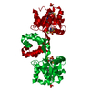
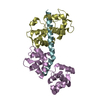
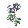
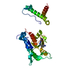
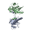
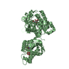
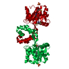
Crystal structure of probable HAD family hydrolase from Pseudomonas fluorescens Pf-5 with bound Mg
Components
Hydrolase, haloacid dehalogenase-like family
Keywords
HYDROLASE / structural genomics / enzyme function intitiative / unknown function / PSI-2 / Protein Structure Initiative / New York SGX Research Center for Structural Genomics / NYSGXRC / haloalkane dehalogenase family / possible hydrolase / Enzyme Function Initiative / EFI
Mass: 18.015 Da / Num. of mol.: 158 / Source method: isolated from a natural source / Formula: H2O
Has protein modification
Y
-
Experimental details
-
Experiment
Experiment
Method: X-RAY DIFFRACTION / Number of used crystals: 1
-
Sample preparation
Crystal
Density Matthews: 3.5 Å3/Da / Density % sol: 64.87 %
Crystal grow
Temperature: 294 K / Method: vapor diffusion, sitting drop / pH: 5.3 Details: Protein 10 mM HEPES pH 8.5, 150 mM Nacl, 10% glycerol, 5 mM DTT, 10 mM MgCl2, Reservoir (2.5 M NaK phosphate pH 5.0, 150 mM NaCitrate), Soak (1.5 M MgSO4, 200 mM MES pH 5.3, 20% glycerol, 10 ...Details: Protein 10 mM HEPES pH 8.5, 150 mM Nacl, 10% glycerol, 5 mM DTT, 10 mM MgCl2, Reservoir (2.5 M NaK phosphate pH 5.0, 150 mM NaCitrate), Soak (1.5 M MgSO4, 200 mM MES pH 5.3, 20% glycerol, 10 min, vapor diffusion, sitting drop, temperature 294K, VAPOR DIFFUSION, SITTING DROP
In the structure databanks used in Yorodumi, some data are registered as the other names, "COVID-19 virus" and "2019-nCoV". Here are the details of the virus and the list of structure data.
Jan 31, 2019. EMDB accession codes are about to change! (news from PDBe EMDB page)
EMDB accession codes are about to change! (news from PDBe EMDB page)
The allocation of 4 digits for EMDB accession codes will soon come to an end. Whilst these codes will remain in use, new EMDB accession codes will include an additional digit and will expand incrementally as the available range of codes is exhausted. The current 4-digit format prefixed with “EMD-” (i.e. EMD-XXXX) will advance to a 5-digit format (i.e. EMD-XXXXX), and so on. It is currently estimated that the 4-digit codes will be depleted around Spring 2019, at which point the 5-digit format will come into force.
The EM Navigator/Yorodumi systems omit the EMD- prefix.
Related info.:Q: What is EMD? / ID/Accession-code notation in Yorodumi/EM Navigator
Yorodumi is a browser for structure data from EMDB, PDB, SASBDB, etc.
This page is also the successor to EM Navigator detail page, and also detail information page/front-end page for Omokage search.
The word "yorodu" (or yorozu) is an old Japanese word meaning "ten thousand". "mi" (miru) is to see.
Related info.:EMDB / PDB / SASBDB / Comparison of 3 databanks / Yorodumi Search / Aug 31, 2016. New EM Navigator & Yorodumi / Yorodumi Papers / Jmol/JSmol / Function and homology information / Changes in new EM Navigator and Yorodumi
 Movie
Movie Controller

 Yorodumi
 Open data
 Basic information
 Components
 Keywords
 Function and homology information
 Pseudomonas fluorescens (bacteria)
 X-RAY DIFFRACTION /
  Authors
 Citation
 Structure visualization
 Downloads & links
 Other downloads


 PDBj
PDBj

 Assembly
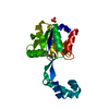

 Mass: 24.305 Da / Num. of mol.: 1 / Source method: obtained synthetically / Formula: Mg
 Mass: 96.063 Da / Num. of mol.: 1 / Source method: obtained synthetically / Formula: SO4
 Mass: 18.015 Da / Num. of mol.: 158 / Source method: isolated from a natural source / Formula: H2O
 Sample preparation
 Processing