Mass: 18.015 Da / Num. of mol.: 229 / Source method: isolated from a natural source / Formula: H2O
Has protein modification
Y
-
Experimental details
-
Experiment
Experiment
Method: X-RAY DIFFRACTION / Number of used crystals: 1
-
Sample preparation
Crystal
Density Matthews: 3.5 Å3/Da / Density % sol: 65 % / Description: NONE
Crystal grow
pH: 5 Details: PROTEIN (10MM HEPES PH 7.5, 150 MM NACL, 10% GLYCEROL, 5 MM DTT, 5 MM MGCL);RESERVOUR (2.5 M NA/K PHOSPHATE, 150 MM NACITRATE, PH 5.0;CRYOPROTECTION (1.8 M NA/K PHOSPHATE, 100 MM NACITRATE, ...Details: PROTEIN (10MM HEPES PH 7.5, 150 MM NACL, 10% GLYCEROL, 5 MM DTT, 5 MM MGCL);RESERVOUR (2.5 M NA/K PHOSPHATE, 150 MM NACITRATE, PH 5.0;CRYOPROTECTION (1.8 M NA/K PHOSPHATE, 100 MM NACITRATE, 18.5% GLYCEROL, 185 MM MGSO4) TWO MINUTE SOAK.
Resolution: 2.001→32.989 Å / SU ML: 0.22 / σ(F): 0 / Phase error: 17.96 / Stereochemistry target values: ML Details: MAGNESIUM IN ACTIVE SITE CHOSEN OVER WATER DUE TO COORDINATION NUMBER AND GEOMETRY. MOST LIKELY DOES NOT REPRESENT THE TYPICAL COORDINATION DURING CHEMISTRY. PHOSPHATE IS ASSUMED TO BE BOUND ...Details: MAGNESIUM IN ACTIVE SITE CHOSEN OVER WATER DUE TO COORDINATION NUMBER AND GEOMETRY. MOST LIKELY DOES NOT REPRESENT THE TYPICAL COORDINATION DURING CHEMISTRY. PHOSPHATE IS ASSUMED TO BE BOUND BY THE ACTIVE SITE POSITION THAT COORDINATES THE PHOSPHATE OF SUBSTRATE.
Rfactor
Num. reflection
% reflection
Rfree
0.2177
1106
5.1 %
Rwork
0.1707
-
-
obs
0.173
21908
95.89 %
Solvent computation
Shrinkage radii: 0.83 Å / VDW probe radii: 1.1 Å / Solvent model: FLAT BULK SOLVENT MODEL / Bsol: 37.422 Å2 / ksol: 0.376 e/Å3
Displacement parameters
Baniso -1
Baniso -2
Baniso -3
1-
0.8479 Å2
0 Å2
0 Å2
2-
-
0.8479 Å2
0 Å2
3-
-
-
-1.6957 Å2
Refinement step
Cycle: LAST / Resolution: 2.001→32.989 Å
Protein
Nucleic acid
Ligand
Solvent
Total
Num. atoms
1507
0
11
229
1747
Refine LS restraints
Refine-ID
Type
Dev ideal
Number
X-RAY DIFFRACTION
f_bond_d
0.008
1591
X-RAY DIFFRACTION
f_angle_d
1.032
2180
X-RAY DIFFRACTION
f_dihedral_angle_d
11.246
582
X-RAY DIFFRACTION
f_chiral_restr
0.071
244
X-RAY DIFFRACTION
f_plane_restr
0.005
288
LS refinement shell
Resolution (Å)
Rfactor Rfree
Num. reflection Rfree
Rfactor Rwork
Num. reflection Rwork
Refine-ID
% reflection obs (%)
2.0009-2.0919
0.2813
125
0.2162
2221
X-RAY DIFFRACTION
85
2.0919-2.2022
0.2101
132
0.1697
2479
X-RAY DIFFRACTION
93
2.2022-2.3401
0.2408
135
0.1659
2523
X-RAY DIFFRACTION
96
2.3401-2.5208
0.2159
137
0.1658
2570
X-RAY DIFFRACTION
97
2.5208-2.7743
0.2093
136
0.1657
2634
X-RAY DIFFRACTION
98
2.7743-3.1755
0.2256
142
0.1653
2680
X-RAY DIFFRACTION
99
3.1755-3.9997
0.1902
145
0.1652
2740
X-RAY DIFFRACTION
100
3.9997-32.9935
0.2237
154
0.1754
2955
X-RAY DIFFRACTION
99
Refinement TLS params.
Method: refined / Refine-ID: X-RAY DIFFRACTION
ID
L11 (°2)
L12 (°2)
L13 (°2)
L22 (°2)
L23 (°2)
L33 (°2)
S11 (Å °)
S12 (Å °)
S13 (Å °)
S21 (Å °)
S22 (Å °)
S23 (Å °)
S31 (Å °)
S32 (Å °)
S33 (Å °)
T11 (Å2)
T12 (Å2)
T13 (Å2)
T22 (Å2)
T23 (Å2)
T33 (Å2)
Origin x (Å)
Origin y (Å)
Origin z (Å)
1
0.2486
0.1412
-0.029
0.3281
-0.0771
0.023
-0.1763
0.1446
-0.0196
-0.0944
0.1445
0.045
-0.043
-0.0538
0.0161
0.1926
0.116
0.0008
-0.0572
0.0444
0.0915
50.4335
6.1734
19.3473
2
0.0946
-0.0174
-0.0544
0.0391
-0.0506
0.1035
-0.1496
-0.009
0.0007
0.0528
0.0713
-0.1307
0.0574
0.0793
0.015
0.1861
0.0172
-0.0424
0.0883
-0.0012
0.1218
65.0061
-7.8637
-2.2223
3
0.0809
-0.0222
-0.0851
0.0229
-0.0167
0.1088
-0.106
-0.0214
-0.0819
0.0501
0.012
0.087
0.1381
-0.0945
0.0088
0.1721
0.0252
0.0057
0.0873
0.0138
0.1114
51.7545
-1.9329
16.8401
4
0.062
-0.0061
0.0126
0.0368
0.0693
0.1984
-0.1554
0.0708
-0.1445
0.0087
-0.0509
0.0786
-0.0135
-0.0377
0.0283
0.1885
0.0469
-0.0098
0.147
-0.0165
0.1604
43.1174
8.0443
20.5064
5
0.1405
-0.1274
-0.0328
0.0998
0.0498
0.1024
-0.064
0.0039
-0.1323
0.0947
0.0068
0.3439
-0.0613
-0.3163
0.0181
0.183
0.0504
-0.0155
0.1019
-0.0257
0.1955
39.1704
8.5432
19.5103
6
0.0898
0.0193
-0.0332
0.0143
-0.0123
0.0648
-0.0168
-0.0659
-0.0314
-0.0678
0.0142
0.0182
-0.2079
0.0285
0.0048
0.1852
0.0498
0.0178
0.0719
0.005
0.088
55.7435
14.1125
18.3558
Refinement TLS group
ID
Refine-ID
Refine TLS-ID
Selection details
1
X-RAY DIFFRACTION
1
CHAIN 'A' AND (RESSEQ2:23)
2
X-RAY DIFFRACTION
2
CHAIN 'A' AND (RESSEQ24:60)
3
X-RAY DIFFRACTION
3
CHAIN 'A' AND (RESSEQ61:85)
4
X-RAY DIFFRACTION
4
CHAIN 'A' AND (RESSEQ86:96)
5
X-RAY DIFFRACTION
5
CHAIN 'A' AND (RESSEQ97:143)
6
X-RAY DIFFRACTION
6
CHAIN 'A' AND (RESSEQ144:197)
+
About Yorodumi
-
News
-
Feb 9, 2022. New format data for meta-information of EMDB entries
New format data for meta-information of EMDB entries
Version 3 of the EMDB header file is now the official format.
The previous official version 1.9 will be removed from the archive.
In the structure databanks used in Yorodumi, some data are registered as the other names, "COVID-19 virus" and "2019-nCoV". Here are the details of the virus and the list of structure data.
Jan 31, 2019. EMDB accession codes are about to change! (news from PDBe EMDB page)
EMDB accession codes are about to change! (news from PDBe EMDB page)
The allocation of 4 digits for EMDB accession codes will soon come to an end. Whilst these codes will remain in use, new EMDB accession codes will include an additional digit and will expand incrementally as the available range of codes is exhausted. The current 4-digit format prefixed with “EMD-” (i.e. EMD-XXXX) will advance to a 5-digit format (i.e. EMD-XXXXX), and so on. It is currently estimated that the 4-digit codes will be depleted around Spring 2019, at which point the 5-digit format will come into force.
The EM Navigator/Yorodumi systems omit the EMD- prefix.
Related info.:Q: What is EMD? / ID/Accession-code notation in Yorodumi/EM Navigator
Yorodumi is a browser for structure data from EMDB, PDB, SASBDB, etc.
This page is also the successor to EM Navigator detail page, and also detail information page/front-end page for Omokage search.
The word "yorodu" (or yorozu) is an old Japanese word meaning "ten thousand". "mi" (miru) is to see.
Related info.:EMDB / PDB / SASBDB / Comparison of 3 databanks / Yorodumi Search / Aug 31, 2016. New EM Navigator & Yorodumi / Yorodumi Papers / Jmol/JSmol / Function and homology information / Changes in new EM Navigator and Yorodumi
 Movie
Movie Controller

 Yorodumi
 Open data
 Basic information
 Components
 Keywords
 Function and homology information
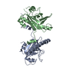
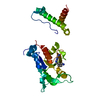
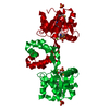
 PSEUDOMONAS FLUORESCENS PF-5 (bacteria)
 X-RAY DIFFRACTION /
  Authors
 Citation
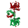
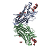
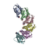
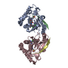
 Structure visualization
 Downloads & links
 Other downloads


 PDBj
PDBj

 Assembly
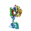

 Mass: 24.305 Da / Num. of mol.: 1 / Source method: obtained synthetically / Formula: Mg
 Mass: 94.971 Da / Num. of mol.: 2 / Source method: obtained synthetically / Formula: PO4
 Mass: 18.015 Da / Num. of mol.: 229 / Source method: isolated from a natural source / Formula: H2O
 Sample preparation
 Processing