 Movie
Movie Controller

[English] 日本語
 Yorodumi
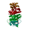
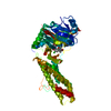
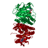
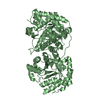
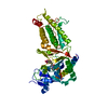
- PDB-3quo: Crystal structure of fosfomycin resistance kinase FomA from Strep... -
+ Open data

- Basic information
| Entry | Database: PDB / ID: 3quo | ||||||
|---|---|---|---|---|---|---|---|
| Title | Crystal structure of fosfomycin resistance kinase FomA from Streptomyces wedmorensis complexed with ATP and fosfomycin | ||||||
 Components Components | FomA protein | ||||||
 Keywords Keywords | TRANSFERASE/TRANSFERASE inhibitor / fosfomycin / antibiotic resistance / phosphoryl transfer / kinase / TRANSFERASE-TRANSFERASE inhibitor complex | ||||||
| Function / homology |  Function and homology information Function and homology informationisopentenyl phosphate kinase / isopentenyl phosphate kinase activity / glutamate 5-kinase activity / isoprenoid biosynthetic process / ATP binding / cytosol Similarity search - Function | ||||||
| Biological species |  Streptomyces wedmorensis (bacteria) Streptomyces wedmorensis (bacteria) | ||||||
| Method |  X-RAY DIFFRACTION / SYNCHROTRON / MOLECULAR REPLACEMENT / Resolution: 1.58 Å X-RAY DIFFRACTION / SYNCHROTRON / MOLECULAR REPLACEMENT / Resolution: 1.58 Å | ||||||
 Authors Authors | Pakhomova, S. / Bartlett, S.G. / Doerner, P.A. / Newcomer, M.E. | ||||||
 Citation Citation | Journal: Biochemistry / Year: 2011 Title: Structural and biochemical insights into the mechanism of fosfomycin phosphorylation by fosfomycin resistance kinase FomA. Authors: Pakhomova, S. / Bartlett, S.G. / Doerner, P.A. / Newcomer, M.E. | ||||||
| History |
|
- Structure visualization
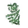
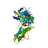
| Structure viewer | Molecule: MolmilJmol/JSmol |
|---|
- Downloads & links
-Download
| PDBx/mmCIF format | 3quo.cif.gz | 121.8 KB | Display | PDBx/mmCIF format |
|---|---|---|---|---|
| PDB format | pdb3quo.ent.gz | 93.3 KB | Display | PDB format |
| PDBx/mmJSON format | 3quo.json.gz | Tree view | PDBx/mmJSON format | |
| Others |  Other downloads Other downloads |
-Validation report
| Arichive directory | https://data.pdbj.org/pub/pdb/validation_reports/qu/3quoftp://data.pdbj.org/pub/pdb/validation_reports/qu/3quo | HTTPS FTP |
|---|
-Related structure data
| Related structure data | 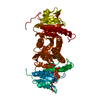 3qunC 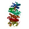 3qurC  3qvfC 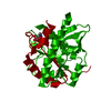 3qvhC 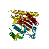 3d40S S: Starting model for refinement C: citing same article ( |
|---|---|
| Similar structure data |


-Links
 PDBj
PDBj- Assembly
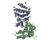
| Deposited unit | 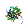
| ||||||||
|---|---|---|---|---|---|---|---|---|---|
| 1 | 
| ||||||||
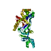
| Unit cell |
|
-Components
| #1: Protein | Mass: 30876.852 Da / Num. of mol.: 1 Source method: isolated from a genetically manipulated source Source: (gene. exp.) Streptomyces wedmorensis (bacteria) / Gene: fomA, fosfomycin resistance kinase / Plasmid: pET28b / Production host: |
|---|---|
| #2: Chemical | ChemComp-ATP /   Mass: 507.181 Da / Num. of mol.: 1 / Source method: obtained synthetically / Formula: C10H16N5O13P3 / Comment: ATP, energy-carrying molecule*YM Mass: 507.181 Da / Num. of mol.: 1 / Source method: obtained synthetically / Formula: C10H16N5O13P3 / Comment: ATP, energy-carrying molecule*YM |
| #3: Chemical | ChemComp-FCN / 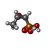  Mass: 138.059 Da / Num. of mol.: 1 / Source method: obtained synthetically / Formula: C3H7O4P / Comment: antibiotic*YM Mass: 138.059 Da / Num. of mol.: 1 / Source method: obtained synthetically / Formula: C3H7O4P / Comment: antibiotic*YM |
| #4: Water | ChemComp-HOH /  Mass: 18.015 Da / Num. of mol.: 203 / Source method: isolated from a natural source / Formula: H2O Mass: 18.015 Da / Num. of mol.: 203 / Source method: isolated from a natural source / Formula: H2O |
| Sequence details | THE AUTHORS STATE THAT THE DEPOSITED SEQUENCE UNPROT ENTRY Q56187 IS WRONG AT POSITION 31 |
-Experimental details
-Experiment
| Experiment | Method: X-RAY DIFFRACTION / Number of used crystals: 1 |
|---|
- Sample preparation
| Crystal | Density Matthews: 2.8 Å3/Da / Density % sol: 56.07 % |
|---|---|
| Crystal grow | Temperature: 298 K / Method: vapor diffusion, hanging drop / pH: 7.1 Details: 17 % PEG3350, 25 % glycerol, 0.1 M MES, 10 mM ATP, 10 mM fosfomycin , pH 7.1, VAPOR DIFFUSION, HANGING DROP, temperature 298K |
-Data collection
| Diffraction | Mean temperature: 100 K |
|---|---|
| Diffraction source | Source: SYNCHROTRON / Site: CAMD  / Beamline: GCPCC / Wavelength: 1.38079 Å / Beamline: GCPCC / Wavelength: 1.38079 Å |
| Detector | Type: MAR CCD 165 mm / Detector: CCD / Date: Jun 10, 2008 / Details: mirrors |
| Radiation | Monochromator: Si 111 channel / Protocol: SINGLE WAVELENGTH / Monochromatic (M) / Laue (L): M / Scattering type: x-ray |
| Radiation wavelength | Wavelength: 1.38079 Å / Relative weight: 1 |
| Reflection | Resolution: 1.58→50 Å / Num. all: 42575 / Num. obs: 42575 / % possible obs: 89 % / Observed criterion σ(I): -3 / Redundancy: 4.4 % / Biso Wilson estimate: 27.4 Å2 / Rsym value: 0.039 / Net I/σ(I): 35.6 |
| Reflection shell | Resolution: 1.58→1.64 Å / Redundancy: 4.2 % / Mean I/σ(I) obs: 1.8 / Num. unique all: 4204 / Rsym value: 0.561 / % possible all: 88.9 |
- Processing
| Software |
| ||||||||||||||||||||||||||||||||||||||||||||||||||||||||||||||||||||||||||||||||||||||||||||||||||||||||||||||||||||||||||||||||||||||||||||||||||||||||||||||||||||||||||
|---|---|---|---|---|---|---|---|---|---|---|---|---|---|---|---|---|---|---|---|---|---|---|---|---|---|---|---|---|---|---|---|---|---|---|---|---|---|---|---|---|---|---|---|---|---|---|---|---|---|---|---|---|---|---|---|---|---|---|---|---|---|---|---|---|---|---|---|---|---|---|---|---|---|---|---|---|---|---|---|---|---|---|---|---|---|---|---|---|---|---|---|---|---|---|---|---|---|---|---|---|---|---|---|---|---|---|---|---|---|---|---|---|---|---|---|---|---|---|---|---|---|---|---|---|---|---|---|---|---|---|---|---|---|---|---|---|---|---|---|---|---|---|---|---|---|---|---|---|---|---|---|---|---|---|---|---|---|---|---|---|---|---|---|---|---|---|---|---|---|---|---|
| Refinement | Method to determine structure: MOLECULAR REPLACEMENT Starting model: pdb entry 3D40 Resolution: 1.58→26.86 Å / Cor.coef. Fo:Fc: 0.97 / Cor.coef. Fo:Fc free: 0.948 / SU B: 2.778 / SU ML: 0.045 / Isotropic thermal model: restrained / Cross valid method: THROUGHOUT / ESU R Free: 0.082 / Stereochemistry target values: MAXIMUM LIKELIHOOD / Details: HYDROGENS HAVE BEEN ADDED IN THE RIDING POSITIONS
| ||||||||||||||||||||||||||||||||||||||||||||||||||||||||||||||||||||||||||||||||||||||||||||||||||||||||||||||||||||||||||||||||||||||||||||||||||||||||||||||||||||||||||
| Solvent computation | Ion probe radii: 0.8 Å / Shrinkage radii: 0.8 Å / VDW probe radii: 1.4 Å / Solvent model: BABINET MODEL WITH MASK | ||||||||||||||||||||||||||||||||||||||||||||||||||||||||||||||||||||||||||||||||||||||||||||||||||||||||||||||||||||||||||||||||||||||||||||||||||||||||||||||||||||||||||
| Displacement parameters | Biso mean: 31.701 Å2
| ||||||||||||||||||||||||||||||||||||||||||||||||||||||||||||||||||||||||||||||||||||||||||||||||||||||||||||||||||||||||||||||||||||||||||||||||||||||||||||||||||||||||||
| Refinement step | Cycle: LAST / Resolution: 1.58→26.86 Å
| ||||||||||||||||||||||||||||||||||||||||||||||||||||||||||||||||||||||||||||||||||||||||||||||||||||||||||||||||||||||||||||||||||||||||||||||||||||||||||||||||||||||||||
| Refine LS restraints |
| ||||||||||||||||||||||||||||||||||||||||||||||||||||||||||||||||||||||||||||||||||||||||||||||||||||||||||||||||||||||||||||||||||||||||||||||||||||||||||||||||||||||||||
| LS refinement shell | Resolution: 1.58→1.621 Å / Total num. of bins used: 20
| ||||||||||||||||||||||||||||||||||||||||||||||||||||||||||||||||||||||||||||||||||||||||||||||||||||||||||||||||||||||||||||||||||||||||||||||||||||||||||||||||||||||||||
| Refinement TLS params. | Method: refined / Refine-ID: X-RAY DIFFRACTION
| ||||||||||||||||||||||||||||||||||||||||||||||||||||||||||||||||||||||||||||||||||||||||||||||||||||||||||||||||||||||||||||||||||||||||||||||||||||||||||||||||||||||||||
| Refinement TLS group |
|
