 Movie
Movie Controller

[English] 日本語
 Yorodumi
- PDB-3qra: The crystal structure of Ail, the attachment invasion locus prote... -
+ Open data

- Basic information
| Entry | Database: PDB / ID: 3qra | ||||||
|---|---|---|---|---|---|---|---|
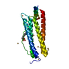
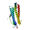
| Title | The crystal structure of Ail, the attachment invasion locus protein of Yersinia pestis | ||||||
 Components Components | Attachment invasion locus protein | ||||||
 Keywords Keywords | CELL INVASION / beta-barrel protein / attachment and invasion virulence / laminin / heparin | ||||||
| Function / homology |  Function and homology information Function and homology information | ||||||
| Biological species |   Yersinia pestis (bacteria) Yersinia pestis (bacteria) | ||||||
| Method |  X-RAY DIFFRACTION / SYNCHROTRON / MOLECULAR REPLACEMENT / Resolution: 1.801 Å X-RAY DIFFRACTION / SYNCHROTRON / MOLECULAR REPLACEMENT / Resolution: 1.801 Å | ||||||
 Authors Authors | Yamashita, S. / Lukacik, P. / Noinaj, N. / Buchanan, S.K. | ||||||
 Citation Citation | Journal: Structure / Year: 2011 Title: Structural Insights into Ail-Mediated Adhesion in Yersinia pestis. Authors: Yamashita, S. / Lukacik, P. / Barnard, T.J. / Noinaj, N. / Felek, S. / Tsang, T.M. / Krukonis, E.S. / Hinnebusch, B.J. / Buchanan, S.K. | ||||||
| History |
|
- Structure visualization
| Structure viewer | Molecule: MolmilJmol/JSmol |
|---|
- Downloads & links
-Download
| PDBx/mmCIF format | 3qra.cif.gz | 77.3 KB | Display | PDBx/mmCIF format |
|---|---|---|---|---|
| PDB format | pdb3qra.ent.gz | 56.7 KB | Display | PDB format |
| PDBx/mmJSON format | 3qra.json.gz | Tree view | PDBx/mmJSON format | |
| Others |  Other downloads Other downloads |
-Validation report
| Summary document | 3qra_validation.pdf.gz | 1.5 MB | Display | wwPDB validaton report |
|---|---|---|---|---|
| Full document | 3qra_full_validation.pdf.gz | 1.5 MB | Display | |
| Data in XML | 3qra_validation.xml.gz | 9.6 KB | Display | |
| Data in CIF | 3qra_validation.cif.gz | 12.3 KB | Display | |
| Arichive directory | https://data.pdbj.org/pub/pdb/validation_reports/qr/3qraftp://data.pdbj.org/pub/pdb/validation_reports/qr/3qra | HTTPS FTP |
-Related structure data
| Related structure data |  3qrcC 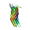 1qj8S C: citing same article ( S: Starting model for refinement |
|---|---|
| Similar structure data |


-Links
 PDBj
PDBj

- Assembly
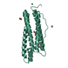
| Deposited unit | 
| ||||||||
|---|---|---|---|---|---|---|---|---|---|
| 1 |
| ||||||||
| Unit cell |
|
-Components
| #1: Protein | Mass: 17623.596 Da / Num. of mol.: 1 / Fragment: UNP residues 27-182 Source method: isolated from a genetically manipulated source Source: (gene. exp.) Yersinia pestis (bacteria) / Gene: ail, YPO2905 / Production host: | ||
|---|---|---|---|
| #2: Chemical | ChemComp-C8E / (   Mass: 306.438 Da / Num. of mol.: 7 / Source method: obtained synthetically / Formula: C16H34O5 / Comment: C8E, detergent*YM Mass: 306.438 Da / Num. of mol.: 7 / Source method: obtained synthetically / Formula: C16H34O5 / Comment: C8E, detergent*YM#3: Water | ChemComp-HOH / |  Mass: 18.015 Da / Num. of mol.: 70 / Source method: isolated from a natural source / Formula: H2O Mass: 18.015 Da / Num. of mol.: 70 / Source method: isolated from a natural source / Formula: H2O |
-Experimental details
-Experiment
| Experiment | Method: X-RAY DIFFRACTION / Number of used crystals: 1 |
|---|
- Sample preparation
| Crystal | Density Matthews: 3.05 Å3/Da / Density % sol: 59.73 % |
|---|---|
| Crystal grow | Temperature: 298 K / Method: vapor diffusion, hanging drop / pH: 6.5 Details: 34% PEG1000, 0.2 M potassium fluoride, 0.1 M Bis-Tris, pH 6.5, VAPOR DIFFUSION, HANGING DROP, temperature 298K |
-Data collection
| Diffraction | Mean temperature: 100 K |
|---|---|
| Diffraction source | Source: SYNCHROTRON / Site: APS  / Beamline: 22-ID / Wavelength: 1 / Beamline: 22-ID / Wavelength: 1 |
| Detector | Type: MARMOSAIC 300 mm CCD / Detector: CCD / Date: Oct 20, 2007 |
| Radiation | Monochromator: double crystal / Protocol: SINGLE WAVELENGTH / Monochromatic (M) / Laue (L): M / Scattering type: x-ray |
| Radiation wavelength | Wavelength: 1 Å / Relative weight: 1 |
| Reflection | Resolution: 1.8→37.7 Å / Num. all: 19652 / Num. obs: 19652 / % possible obs: 93 % / Observed criterion σ(F): 1 / Observed criterion σ(I): 1 / Redundancy: 12.3 % / Rsym value: 0.058 / Net I/σ(I): 44.1 |
| Reflection shell | Resolution: 1.8→1.86 Å / Redundancy: 4.7 % / Mean I/σ(I) obs: 3.9 / Rsym value: 0.346 / % possible all: 68 |
- Processing
| Software |
| ||||||||||||||||||||||||||||||||||||||||||||||||||||||||
|---|---|---|---|---|---|---|---|---|---|---|---|---|---|---|---|---|---|---|---|---|---|---|---|---|---|---|---|---|---|---|---|---|---|---|---|---|---|---|---|---|---|---|---|---|---|---|---|---|---|---|---|---|---|---|---|---|---|
| Refinement | Method to determine structure: MOLECULAR REPLACEMENT Starting model: PDB ENTRY 1QJ8 Resolution: 1.801→34.777 Å / SU ML: 0.2 / σ(F): 0 / Phase error: 20.13 / Stereochemistry target values: ML
| ||||||||||||||||||||||||||||||||||||||||||||||||||||||||
| Solvent computation | Shrinkage radii: 0.72 Å / VDW probe radii: 1 Å / Solvent model: FLAT BULK SOLVENT MODEL / Bsol: 80 Å2 / ksol: 0.4 e/Å3 | ||||||||||||||||||||||||||||||||||||||||||||||||||||||||
| Displacement parameters |
| ||||||||||||||||||||||||||||||||||||||||||||||||||||||||
| Refinement step | Cycle: LAST / Resolution: 1.801→34.777 Å
| ||||||||||||||||||||||||||||||||||||||||||||||||||||||||
| Refine LS restraints |
| ||||||||||||||||||||||||||||||||||||||||||||||||||||||||
| LS refinement shell |
| ||||||||||||||||||||||||||||||||||||||||||||||||||||||||
| Refinement TLS params. | Method: refined / Origin x: 8.6544 Å / Origin y: 19.8181 Å / Origin z: 24.9946 Å
| ||||||||||||||||||||||||||||||||||||||||||||||||||||||||
| Refinement TLS group | Selection details: chain A |
