 Movie
Movie Controller

[English] 日本語
 Yorodumi
- PDB-1qj8: CRYSTAL STRUCTURE OF THE OUTER MEMBRANE PROTEIN OMPX FROM ESCHERI... -
+ Open data

- Basic information
| Entry | Database: PDB / ID: 1qj8 | ||||||
|---|---|---|---|---|---|---|---|
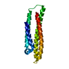
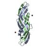
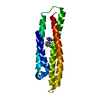
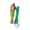
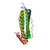
| Title | CRYSTAL STRUCTURE OF THE OUTER MEMBRANE PROTEIN OMPX FROM ESCHERICHIA COLI | ||||||
 Components Components | OUTER MEMBRANE PROTEIN X | ||||||
 Keywords Keywords | MEMBRANE PROTEIN / BETA-BARREL / BACTERIAL DEFENSE SYSTEM / PLATINUM COMPLEX STRUCTURE | ||||||
| Function / homology |  Function and homology information Function and homology information | ||||||
| Biological species |  | ||||||
| Method |  X-RAY DIFFRACTION / SYNCHROTRON / MIR / Resolution: 1.9 Å X-RAY DIFFRACTION / SYNCHROTRON / MIR / Resolution: 1.9 Å | ||||||
 Authors Authors | Vogt, J. / Schulz, G.E. | ||||||
 Citation Citation | Journal: Structure / Year: 1999 Title: The Structure of the Outer Membrane Protein Ompx from Escherichia Coli Reveals Mechanisms of Virulence Authors: Vogt, J. / Schulz, G.E. #1: Journal: Proteins / Year: 1999 Title: Strategy for Membrane Protein Crystallization Exemplified with Ompa and Ompx Authors: Pautsch, A. / Vogt, J. / Model, K. / Siebold, C. / Schulz, G.E. | ||||||
| History |
| ||||||
| Remark 700 | SHEET DETERMINATION METHOD: DSSP THE SHEET PRESENTED AS "A" ON SHEET RECORDS BELOW IS ACTUALLY AN ... SHEET DETERMINATION METHOD: DSSP THE SHEET PRESENTED AS "A" ON SHEET RECORDS BELOW IS ACTUALLY AN 8-STRANDED BETA-BARREL. THIS IS REPRESENTED BY A 9-STRANDED SHEET IN WHICH THE FIRST AND LAST STRANDS ARE IDENTICAL. |
- Structure visualization
| Structure viewer | Molecule: MolmilJmol/JSmol |
|---|
- Downloads & links
-Download
| PDBx/mmCIF format | 1qj8.cif.gz | 45.6 KB | Display | PDBx/mmCIF format |
|---|---|---|---|---|
| PDB format | pdb1qj8.ent.gz | 32.6 KB | Display | PDB format |
| PDBx/mmJSON format | 1qj8.json.gz | Tree view | PDBx/mmJSON format | |
| Others |  Other downloads Other downloads |
-Validation report
| Arichive directory | https://data.pdbj.org/pub/pdb/validation_reports/qj/1qj8ftp://data.pdbj.org/pub/pdb/validation_reports/qj/1qj8 | HTTPS FTP |
|---|
-Related structure data


-Links
 PDBj
PDBj

- Assembly
| Deposited unit | 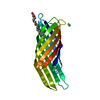
| ||||||||
|---|---|---|---|---|---|---|---|---|---|
| 1 |
| ||||||||
| Unit cell |
| ||||||||
| Components on special symmetry positions |
|
-Components
| #1: Protein | Mass: 16371.768 Da / Num. of mol.: 1 / Mutation: YES Source method: isolated from a genetically manipulated source Source: (gene. exp.) | ||||
|---|---|---|---|---|---|
| #2: Chemical | ChemComp-C8E / (  Mass: 306.438 Da / Num. of mol.: 1 / Source method: obtained synthetically / Formula: C16H34O5 / Comment: C8E, detergent*YM Mass: 306.438 Da / Num. of mol.: 1 / Source method: obtained synthetically / Formula: C16H34O5 / Comment: C8E, detergent*YM | ||||
| #3: Chemical |   Mass: 265.984 Da / Num. of mol.: 2 / Source method: obtained synthetically / Formula: Cl2Pt Mass: 265.984 Da / Num. of mol.: 2 / Source method: obtained synthetically / Formula: Cl2Pt#4: Water | ChemComp-HOH / |  Mass: 18.015 Da / Num. of mol.: 72 / Source method: isolated from a natural source / Formula: H2O Mass: 18.015 Da / Num. of mol.: 72 / Source method: isolated from a natural source / Formula: H2OCompound details | ENGINEERED | |
-Experimental details
-Experiment
| Experiment | Method: X-RAY DIFFRACTION / Number of used crystals: 1 |
|---|
- Sample preparation
| Crystal | Density Matthews: 4.1 Å3/Da / Density % sol: 70 % | ||||||||||||||||||||||||||||||||||||||||||||||||
|---|---|---|---|---|---|---|---|---|---|---|---|---|---|---|---|---|---|---|---|---|---|---|---|---|---|---|---|---|---|---|---|---|---|---|---|---|---|---|---|---|---|---|---|---|---|---|---|---|---|
| Crystal grow | pH: 4.6 Details: 30 % (V/V) 2-PROPANOL, 20 % (V/V) GLYCEROL, 0.2 M CACL2, 0.1 M NA-ACETATE PH 4.6 | ||||||||||||||||||||||||||||||||||||||||||||||||
| Crystal | *PLUS Density % sol: 70 % | ||||||||||||||||||||||||||||||||||||||||||||||||
| Crystal grow | *PLUS pH: 8.5 / Method: vapor diffusion, hanging drop | ||||||||||||||||||||||||||||||||||||||||||||||||
| Components of the solutions | *PLUS
|
-Data collection
| Diffraction | Mean temperature: 100 K |
|---|---|
| Diffraction source | Source: SYNCHROTRON / Site: EMBL/DESY, HAMBURG  / Beamline: X11 / Wavelength: 0.9057 / Beamline: X11 / Wavelength: 0.9057 |
| Detector | Type: MARRESEARCH / Detector: IMAGE PLATE / Date: Apr 15, 1998 |
| Radiation | Protocol: SINGLE WAVELENGTH / Monochromatic (M) / Laue (L): M / Scattering type: x-ray |
| Radiation wavelength | Wavelength: 0.9057 Å / Relative weight: 1 |
| Reflection | Resolution: 1.9→34 Å / Num. obs: 20466 / % possible obs: 95 % / Observed criterion σ(I): 0 / Redundancy: 3.7 % / Biso Wilson estimate: 27 Å2 / Rsym value: 0.061 / Net I/σ(I): 14.6 |
| Reflection shell | Resolution: 1.9→2 Å / Redundancy: 3 % / Mean I/σ(I) obs: 2.9 / Rsym value: 0.274 / % possible all: 92 |
| Reflection | *PLUS Rmerge(I) obs: 0.061 |
| Reflection shell | *PLUS Rmerge(I) obs: 0.27 |
- Processing
| Software |
| |||||||||||||||||||||||||||||||||||||||||||||||||||||||||||||||
|---|---|---|---|---|---|---|---|---|---|---|---|---|---|---|---|---|---|---|---|---|---|---|---|---|---|---|---|---|---|---|---|---|---|---|---|---|---|---|---|---|---|---|---|---|---|---|---|---|---|---|---|---|---|---|---|---|---|---|---|---|---|---|---|---|
| Refinement | Method to determine structure: MIR / Resolution: 1.9→34 Å / SU B: 1.539 / SU ML: 0.046 / Cross valid method: THROUGHOUT / σ(F): 0 / ESU R: 0.125 / ESU R Free: 0.128
| |||||||||||||||||||||||||||||||||||||||||||||||||||||||||||||||
| Displacement parameters | Biso mean: 44 Å2 | |||||||||||||||||||||||||||||||||||||||||||||||||||||||||||||||
| Refinement step | Cycle: LAST / Resolution: 1.9→34 Å
| |||||||||||||||||||||||||||||||||||||||||||||||||||||||||||||||
| Refine LS restraints |
| |||||||||||||||||||||||||||||||||||||||||||||||||||||||||||||||
| Software | *PLUS Name: REFMAC / Classification: refinement | |||||||||||||||||||||||||||||||||||||||||||||||||||||||||||||||
| Refinement | *PLUS Rfactor obs: 0.204 | |||||||||||||||||||||||||||||||||||||||||||||||||||||||||||||||
| Solvent computation | *PLUS | |||||||||||||||||||||||||||||||||||||||||||||||||||||||||||||||
| Displacement parameters | *PLUS |
