 Movie
Movie Controller


+ Open data

- Basic information
| Entry | Database: PDB / ID: 3qnw | ||||||
|---|---|---|---|---|---|---|---|
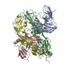
| Title | Caspase-6 in complex with Z-VAD-FMK inhibitor | ||||||
 Components Components |
| ||||||
 Keywords Keywords | HYDROLASE/HYDROLASE INHIBITOR / cysteine protease / HYDROLASE-HYDROLASE INHIBITOR complex | ||||||
| Function / homology |  Function and homology information Function and homology informationcaspase-6 / Breakdown of the nuclear lamina / regulation of programmed cell death / cellular response to staurosporine / positive regulation of necroptotic process / Apoptotic cleavage of cellular proteins / TP53 Regulates Transcription of Caspase Activators and Caspases / hepatocyte apoptotic process / protein autoprocessing / intrinsic apoptotic signaling pathway by p53 class mediator ...caspase-6 / Breakdown of the nuclear lamina / regulation of programmed cell death / cellular response to staurosporine / positive regulation of necroptotic process / Apoptotic cleavage of cellular proteins / TP53 Regulates Transcription of Caspase Activators and Caspases / hepatocyte apoptotic process / protein autoprocessing / intrinsic apoptotic signaling pathway by p53 class mediator / pyroptotic inflammatory response / Caspase-mediated cleavage of cytoskeletal proteins / epithelial cell differentiation / cysteine-type peptidase activity / activation of innate immune response / positive regulation of neuron apoptotic process / positive regulation of apoptotic process / cysteine-type endopeptidase activity / apoptotic process / proteolysis / nucleoplasm / identical protein binding / nucleus / cytoplasm / cytosol Similarity search - Function | ||||||
| Biological species |  Homo sapiens (human) Homo sapiens (human) | ||||||
| Method |  X-RAY DIFFRACTION / SYNCHROTRON / MOLECULAR REPLACEMENT / Resolution: 2.65 Å X-RAY DIFFRACTION / SYNCHROTRON / MOLECULAR REPLACEMENT / Resolution: 2.65 Å | ||||||
 Authors Authors | Mueller, I. / Lamers, M. / Ritchie, A. / Park, H. / Dominguez, C. / Munoz, I. / Maillard, M. / Kiselyov, A. | ||||||
 Citation Citation | Journal: Bioorg.Med.Chem.Lett. / Year: 2011 Title: Structure of human caspase-6 in complex with Z-VAD-FMK: New peptide binding mode observed for the non-canonical caspase conformation. Authors: Muller, I. / Lamers, M.B. / Ritchie, A.J. / Dominguez, C. / Munoz-Sanjuan, I. / Kiselyov, A. | ||||||
| History |
|
- Structure visualization
| Structure viewer | Molecule: MolmilJmol/JSmol |
|---|
- Downloads & links
-Download
| PDBx/mmCIF format | 3qnw.cif.gz | 181.9 KB | Display | PDBx/mmCIF format |
|---|---|---|---|---|
| PDB format | pdb3qnw.ent.gz | 143 KB | Display | PDB format |
| PDBx/mmJSON format | 3qnw.json.gz | Tree view | PDBx/mmJSON format | |
| Others |  Other downloads Other downloads |
-Validation report
| Arichive directory | https://data.pdbj.org/pub/pdb/validation_reports/qn/3qnwftp://data.pdbj.org/pub/pdb/validation_reports/qn/3qnw | HTTPS FTP |
|---|
-Related structure data
| Related structure data |  2wdpS S: Starting model for refinement |
|---|---|
| Similar structure data |


-Links
 PDBj
PDBj


- Assembly
| Deposited unit | 
| ||||||||
|---|---|---|---|---|---|---|---|---|---|
| 1 | 
| ||||||||
| 2 | 
| ||||||||
| Unit cell |
| ||||||||
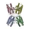
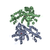
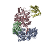
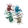
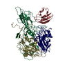
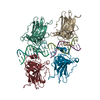
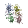
| Details | PROTEIN FORMS DIMERS OF DIMERS (CHAINS A/B AND C/D AND CHAINS E/F AND G/H). LIGAND DOES NOT BIND TO ALL HETERODIMERS |
-Components
| #1: Protein | Mass: 18098.809 Da / Num. of mol.: 4 / Fragment: unp residues 24-179 Source method: isolated from a genetically manipulated source Details: caspase-6 zymogen expressed in E.coli, mature caspase-6 via autoproteolysis Source: (gene. exp.) Homo sapiens (human) / Gene: CASP6, MCH2 / Production host:  #2: Protein | Mass: 11300.012 Da / Num. of mol.: 4 / Fragment: unp residues 194-293 Source method: isolated from a genetically manipulated source Details: caspase-6 zymogen expressed in E.coli, mature caspase-6 via autoproteolysis Source: (gene. exp.) Homo sapiens (human) / Gene: CASP6, MCH2 / Production host: #3: Protein/peptide |   Type: Peptide-like / Class: Inhibitor / Mass: 471.907 Da / Num. of mol.: 3 / Source method: obtained synthetically Type: Peptide-like / Class: Inhibitor / Mass: 471.907 Da / Num. of mol.: 3 / Source method: obtained syntheticallyReferences: N-[(benzyloxy)carbonyl]-L-valyl-N-[(1S)-1-(carboxymethyl)-3-fluoro-2-oxopropyl]-L-alaninamide #4: Water | ChemComp-HOH / |  Mass: 18.015 Da / Num. of mol.: 80 / Source method: isolated from a natural source / Formula: H2O Mass: 18.015 Da / Num. of mol.: 80 / Source method: isolated from a natural source / Formula: H2OHas protein modification | Y | |
|---|
-Experimental details
-Experiment
| Experiment | Method: X-RAY DIFFRACTION / Number of used crystals: 1 |
|---|
- Sample preparation
| Crystal | Density Matthews: 2.2 Å3/Da / Density % sol: 44.06 % |
|---|---|
| Crystal grow | Temperature: 298 K / Method: vapor diffusion / pH: 4.3 Details: 0.1 M lithium sulfate, 0.1 M sodium citrate, 11% 2-propanol , VAPOR DIFFUSION, temperature 298K, pH 4.3 |
-Data collection
| Diffraction | Mean temperature: 100 K |
|---|---|
| Diffraction source | Source: SYNCHROTRON / Site: Diamond  / Beamline: I04 / Wavelength: 0.9763 Å / Beamline: I04 / Wavelength: 0.9763 Å |
| Detector | Type: ADSC QUANTUM 315 / Detector: CCD / Date: Jan 1, 2010 |
| Radiation | Protocol: SINGLE WAVELENGTH / Monochromatic (M) / Laue (L): M / Scattering type: x-ray |
| Radiation wavelength | Wavelength: 0.9763 Å / Relative weight: 1 |
| Reflection | Resolution: 2.65→29.1 Å / Num. all: 30244 / Num. obs: 30002 / % possible obs: 99.2 % / Redundancy: 7.5 % / Rmerge(I) obs: 0.114 / Net I/σ(I): 14.8 |
| Reflection shell | Resolution: 2.65→2.79 Å / Redundancy: 7.6 % / Rmerge(I) obs: 0.564 / Mean I/σ(I) obs: 3.6 / % possible all: 98.8 |
- Processing
| Software |
| ||||||||||||||||||||||||||||||||||||||||||||||||||||||||||||||||||||||||||||||||||||||||||||||||||||||||||||||||||||||||||||||||||||||||||||||||||||||||||||||||||||||||||
|---|---|---|---|---|---|---|---|---|---|---|---|---|---|---|---|---|---|---|---|---|---|---|---|---|---|---|---|---|---|---|---|---|---|---|---|---|---|---|---|---|---|---|---|---|---|---|---|---|---|---|---|---|---|---|---|---|---|---|---|---|---|---|---|---|---|---|---|---|---|---|---|---|---|---|---|---|---|---|---|---|---|---|---|---|---|---|---|---|---|---|---|---|---|---|---|---|---|---|---|---|---|---|---|---|---|---|---|---|---|---|---|---|---|---|---|---|---|---|---|---|---|---|---|---|---|---|---|---|---|---|---|---|---|---|---|---|---|---|---|---|---|---|---|---|---|---|---|---|---|---|---|---|---|---|---|---|---|---|---|---|---|---|---|---|---|---|---|---|---|---|---|
| Refinement | Method to determine structure: MOLECULAR REPLACEMENT Starting model: pdb entry 2wdp Resolution: 2.65→29.1 Å / Cor.coef. Fo:Fc: 0.896 / Cor.coef. Fo:Fc free: 0.856 / SU B: 13.724 / SU ML: 0.292 / Cross valid method: THROUGHOUT / ESU R: 2.515 / ESU R Free: 0.386 / Stereochemistry target values: MAXIMUM LIKELIHOOD / Details: HYDROGENS HAVE BEEN ADDED IN THE RIDING POSITIONS
| ||||||||||||||||||||||||||||||||||||||||||||||||||||||||||||||||||||||||||||||||||||||||||||||||||||||||||||||||||||||||||||||||||||||||||||||||||||||||||||||||||||||||||
| Solvent computation | Ion probe radii: 0.8 Å / Shrinkage radii: 0.8 Å / VDW probe radii: 1.4 Å / Solvent model: MASK | ||||||||||||||||||||||||||||||||||||||||||||||||||||||||||||||||||||||||||||||||||||||||||||||||||||||||||||||||||||||||||||||||||||||||||||||||||||||||||||||||||||||||||
| Displacement parameters | Biso mean: 24.792 Å2
| ||||||||||||||||||||||||||||||||||||||||||||||||||||||||||||||||||||||||||||||||||||||||||||||||||||||||||||||||||||||||||||||||||||||||||||||||||||||||||||||||||||||||||
| Refinement step | Cycle: LAST / Resolution: 2.65→29.1 Å
| ||||||||||||||||||||||||||||||||||||||||||||||||||||||||||||||||||||||||||||||||||||||||||||||||||||||||||||||||||||||||||||||||||||||||||||||||||||||||||||||||||||||||||
| Refine LS restraints |
| ||||||||||||||||||||||||||||||||||||||||||||||||||||||||||||||||||||||||||||||||||||||||||||||||||||||||||||||||||||||||||||||||||||||||||||||||||||||||||||||||||||||||||
| LS refinement shell | Resolution: 2.65→2.719 Å / Total num. of bins used: 20
|
