 Movie
Movie Controller

[English] 日本語
 Yorodumi
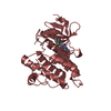
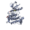
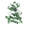
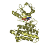
- PDB-3qlf: Crystal structure of the L317I mutant of the C-src tyrosine kinas... -
+ Open data

- Basic information
| Entry | Database: PDB / ID: 3qlf | ||||||
|---|---|---|---|---|---|---|---|
| Title | Crystal structure of the L317I mutant of the C-src tyrosine kinase domain complexed with pyrazolopyrimidine 5 | ||||||
 Components Components | Proto-oncogene tyrosine-protein kinase Src | ||||||
 Keywords Keywords | TRANSFERASE / C-SRC L317I MUTANT / TYROSINE KINASE / PYRAZOLOPYRIMIDINE 5 | ||||||
| Function / homology |  Function and homology information Function and homology informationSignaling by ERBB2 / Nuclear signaling by ERBB4 / Signaling by SCF-KIT / Regulation of KIT signaling / Signaling by EGFR / GAB1 signalosome / Regulation of gap junction activity / FCGR activation / PECAM1 interactions / Co-stimulation by CD28 ...Signaling by ERBB2 / Nuclear signaling by ERBB4 / Signaling by SCF-KIT / Regulation of KIT signaling / Signaling by EGFR / GAB1 signalosome / Regulation of gap junction activity / FCGR activation / PECAM1 interactions / Co-stimulation by CD28 / Co-inhibition by CTLA4 / EPHA-mediated growth cone collapse / Ephrin signaling / G alpha (i) signalling events / GP1b-IX-V activation signalling / Thrombin signalling through proteinase activated receptors (PARs) / VEGFR2 mediated cell proliferation / RET signaling / Receptor Mediated Mitophagy / : / ADP signalling through P2Y purinoceptor 1 / RAF activation / PIP3 activates AKT signaling / EPH-ephrin mediated repulsion of cells / PI5P, PP2A and IER3 Regulate PI3K/AKT Signaling / Downstream signal transduction / Downregulation of ERBB4 signaling / Cyclin D associated events in G1 / Regulation of RUNX3 expression and activity / Degradation of CDH1 / MAP2K and MAPK activation / Integrin signaling / GRB2:SOS provides linkage to MAPK signaling for Integrins / : / MET activates PTK2 signaling / Extra-nuclear estrogen signaling / EPHB-mediated forward signaling / p130Cas linkage to MAPK signaling for integrins / VEGFA-VEGFR2 Pathway / connexin binding / negative regulation of intrinsic apoptotic signaling pathway / progesterone receptor signaling pathway / immune system process / negative regulation of extrinsic apoptotic signaling pathway / non-specific protein-tyrosine kinase / non-membrane spanning protein tyrosine kinase activity / epidermal growth factor receptor signaling pathway / cell junction / cell-cell junction / protein tyrosine kinase activity / protein phosphatase binding / cytoskeleton / cell differentiation / cell adhesion / regulation of cell cycle / endosome membrane / mitochondrial inner membrane / signaling receptor binding / focal adhesion / heme binding / perinuclear region of cytoplasm / protein-containing complex / ATP binding / membrane / nucleus / plasma membrane / cytosol Similarity search - Function | ||||||
| Biological species |  | ||||||
| Method |  X-RAY DIFFRACTION / SYNCHROTRON / MOLECULAR REPLACEMENT / Resolution: 2.75 Å X-RAY DIFFRACTION / SYNCHROTRON / MOLECULAR REPLACEMENT / Resolution: 2.75 Å | ||||||
 Authors Authors | Boubeva, R. / Pernot, L. / Perozzo, R. / Scapozza, L. | ||||||
 Citation Citation | Journal: To be Published Title: A single amino-acid dictates the dynamics of the switch between active and inactive C-src conformation Authors: Boubeva, R. / Pernot, L. / Cristiani, A. / Moretti, L. / Berteotti, A. / Perozzo, R. / Gervasio, F. / Scapozza, L. #1: Journal: Structure / Year: 2007Title: c-Src binds to the cancer drug imatinib with an inactive Abl/c-Kit conformation and a distributed thermodynamic penalty. Authors: Seeliger, M.A. / Nagar, B. / Frank, F. / Cao, X. / Henderson, M.N. / Kuriyan, J. #2: Journal: Chem.Biol. / Year: 2008Title: Small molecule recognition of c-Src via the Imatinib-binding conformation. Authors: Dar, A.C. / Lopez, M.S. / Shokat, K.M. | ||||||
| History |
|
- Structure visualization
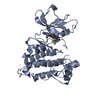
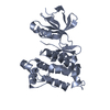
| Structure viewer | Molecule: MolmilJmol/JSmol |
|---|
- Downloads & links
-Download
| PDBx/mmCIF format | 3qlf.cif.gz | 118.9 KB | Display | PDBx/mmCIF format |
|---|---|---|---|---|
| PDB format | pdb3qlf.ent.gz | 91.2 KB | Display | PDB format |
| PDBx/mmJSON format | 3qlf.json.gz | Tree view | PDBx/mmJSON format | |
| Others |  Other downloads Other downloads |
-Validation report
| Arichive directory | https://data.pdbj.org/pub/pdb/validation_reports/ql/3qlfftp://data.pdbj.org/pub/pdb/validation_reports/ql/3qlf | HTTPS FTP |
|---|
-Related structure data
| Related structure data | 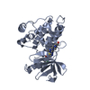 3qlgC 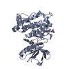 3oezS S: Starting model for refinement C: citing same article ( |
|---|---|
| Similar structure data |


-Links
 PDBj
PDBj- Assembly
| Deposited unit | 
| |||||||||||||||||||||||||||||||||||||||||||||||||||||||||||
|---|---|---|---|---|---|---|---|---|---|---|---|---|---|---|---|---|---|---|---|---|---|---|---|---|---|---|---|---|---|---|---|---|---|---|---|---|---|---|---|---|---|---|---|---|---|---|---|---|---|---|---|---|---|---|---|---|---|---|---|---|
| 1 | 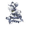
| |||||||||||||||||||||||||||||||||||||||||||||||||||||||||||
| 2 | 
| |||||||||||||||||||||||||||||||||||||||||||||||||||||||||||
| Unit cell |
| |||||||||||||||||||||||||||||||||||||||||||||||||||||||||||
| Noncrystallographic symmetry (NCS) | NCS domain:
NCS domain segments:
NCS ensembles :
|
-Components
| #1: Protein | Mass: 32726.645 Da / Num. of mol.: 2 / Mutation: L317I Source method: isolated from a genetically manipulated source Source: (gene. exp.)  References: UniProt: P00523, non-specific protein-tyrosine kinase #2: Chemical |   Mass: 455.436 Da / Num. of mol.: 2 / Source method: obtained synthetically / Formula: C22H20F3N7O Mass: 455.436 Da / Num. of mol.: 2 / Source method: obtained synthetically / Formula: C22H20F3N7O#3: Water | ChemComp-HOH / |  Mass: 18.015 Da / Num. of mol.: 110 / Source method: isolated from a natural source / Formula: H2O Mass: 18.015 Da / Num. of mol.: 110 / Source method: isolated from a natural source / Formula: H2O |
|---|
-Experimental details
-Experiment
| Experiment | Method: X-RAY DIFFRACTION / Number of used crystals: 1 |
|---|
- Sample preparation
| Crystal | Density Matthews: 2.96 Å3/Da / Density % sol: 58.46 % |
|---|---|
| Crystal grow | Temperature: 287 K / Method: vapor diffusion, hanging drop / pH: 6.5 Details: 7mg/ml cSRcL317I, 0.5mM pyrazolopyrimidine 5, 0.1M MES, 0.2M sodium acetate, 4% glycerol, 12% PEG 4000, pH 6.5, VAPOR DIFFUSION, HANGING DROP, temperature 287K |
-Data collection
| Diffraction | Mean temperature: 100 K |
|---|---|
| Diffraction source | Source: SYNCHROTRON / Site: SLS  / Beamline: X06DA / Wavelength: 1 Å / Beamline: X06DA / Wavelength: 1 Å |
| Detector | Type: MARMOSAIC 225 mm CCD / Detector: CCD / Date: Apr 28, 2009 |
| Radiation | Protocol: SINGLE WAVELENGTH / Monochromatic (M) / Laue (L): M / Scattering type: x-ray |
| Radiation wavelength | Wavelength: 1 Å / Relative weight: 1 |
| Reflection | Resolution: 2.75→32.945 Å / Num. obs: 18749 / % possible obs: 96 % / Observed criterion σ(I): 2 / Redundancy: 3.9 % / Biso Wilson estimate: 52.9 Å2 / Rmerge(I) obs: 0.165 / Rsym value: 0.117 / Net I/σ(I): 10.3 |
| Reflection shell | Resolution: 2.75→2.9 Å / Redundancy: 3.9 % / Rmerge(I) obs: 0.761 / Mean I/σ(I) obs: 2.22 / Rsym value: 0.538 / % possible all: 95.2 |
- Processing
| Software |
| |||||||||||||||||||||||||||||||||||||||||||||||||||||||||||||||
|---|---|---|---|---|---|---|---|---|---|---|---|---|---|---|---|---|---|---|---|---|---|---|---|---|---|---|---|---|---|---|---|---|---|---|---|---|---|---|---|---|---|---|---|---|---|---|---|---|---|---|---|---|---|---|---|---|---|---|---|---|---|---|---|---|
| Refinement | Method to determine structure: MOLECULAR REPLACEMENT Starting model: PDB entry 3OEZ Resolution: 2.75→32.945 Å / SU ML: 0.35 / σ(F): 2.07 / Phase error: 21.48 / Stereochemistry target values: ML
| |||||||||||||||||||||||||||||||||||||||||||||||||||||||||||||||
| Solvent computation | Shrinkage radii: 0.72 Å / VDW probe radii: 1 Å / Solvent model: FLAT BULK SOLVENT MODEL / Bsol: 39.249 Å2 / ksol: 0.352 e/Å3 | |||||||||||||||||||||||||||||||||||||||||||||||||||||||||||||||
| Displacement parameters | Biso mean: 39.21 Å2
| |||||||||||||||||||||||||||||||||||||||||||||||||||||||||||||||
| Refinement step | Cycle: LAST / Resolution: 2.75→32.945 Å
| |||||||||||||||||||||||||||||||||||||||||||||||||||||||||||||||
| Refine LS restraints |
| |||||||||||||||||||||||||||||||||||||||||||||||||||||||||||||||
| Refine LS restraints NCS |
| |||||||||||||||||||||||||||||||||||||||||||||||||||||||||||||||
| LS refinement shell |
|
