 Movie
Movie Controller

[English] 日本語
 Yorodumi
- PDB-3q9n: In silico and in vitro co-evolution of a high affinity complement... -
+ Open data

- Basic information
| Entry | Database: PDB / ID: 3q9n | ||||||
|---|---|---|---|---|---|---|---|
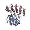
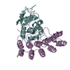
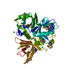
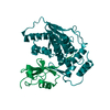
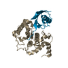
| Title | In silico and in vitro co-evolution of a high affinity complementary protein-protein interface | ||||||
 Components Components |
| ||||||
 Keywords Keywords | PROTEIN BINDING / DE NOVO PROTEIN / Structural Genomics / Israel Structural Proteomics Center / ISPC / Prb-binding designed ankyrin repeat | ||||||
| Function / homology |  Function and homology information Function and homology informationAnkyrin repeat-containing domain / Serine Threonine Protein Phosphatase 5, Tetratricopeptide repeat / Alpha Horseshoe / NAD(P)-binding Rossmann-like Domain / Rossmann fold / 3-Layer(aba) Sandwich / Mainly Alpha / Alpha Beta Similarity search - Domain/homology | ||||||
| Biological species |  | ||||||
| Method |  X-RAY DIFFRACTION / SYNCHROTRON / MOLECULAR REPLACEMENT / Resolution: 2 Å X-RAY DIFFRACTION / SYNCHROTRON / MOLECULAR REPLACEMENT / Resolution: 2 Å | ||||||
 Authors Authors | Karanicolas, J. / Corn, J.E. / Chen, I. / Joachimiak, L.A. / Dym, O. / Chung, S. / Albeck, S. / Unger, T. / Hu, W. / Liu, G. ...Karanicolas, J. / Corn, J.E. / Chen, I. / Joachimiak, L.A. / Dym, O. / Chung, S. / Albeck, S. / Unger, T. / Hu, W. / Liu, G. / Delbecq, S. / Montelione, G.T. / Spiegel, C. / Liu, D. / Baker, D. / Israel Structural Proteomics Center (ISPC) | ||||||
 Citation Citation | Journal: Mol.Cell / Year: 2011 Title: A de novo protein binding pair by computational design and directed evolution. Authors: Karanicolas, J. / Corn, J.E. / Chen, I. / Joachimiak, L.A. / Dym, O. / Peck, S.H. / Albeck, S. / Unger, T. / Hu, W. / Liu, G. / Delbecq, S. / Montelione, G.T. / Spiegel, C.P. / Liu, D.R. / Baker, D. | ||||||
| History |
|
- Structure visualization
| Structure viewer | Molecule: MolmilJmol/JSmol |
|---|
- Downloads & links
-Download
| PDBx/mmCIF format | 3q9n.cif.gz | 243.9 KB | Display | PDBx/mmCIF format |
|---|---|---|---|---|
| PDB format | pdb3q9n.ent.gz | 195.7 KB | Display | PDB format |
| PDBx/mmJSON format | 3q9n.json.gz | Tree view | PDBx/mmJSON format | |
| Others |  Other downloads Other downloads |
-Validation report
| Arichive directory | https://data.pdbj.org/pub/pdb/validation_reports/q9/3q9nftp://data.pdbj.org/pub/pdb/validation_reports/q9/3q9n | HTTPS FTP |
|---|
-Related structure data
| Related structure data |  3q9uC 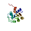 3qa9C 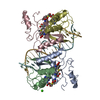 1mjoS 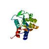 2d59S C: citing same article ( S: Starting model for refinement |
|---|---|
| Similar structure data |


-Links
 PDBj
PDBj


- Assembly
| Deposited unit | 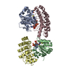
| ||||||||
|---|---|---|---|---|---|---|---|---|---|
| 1 | 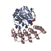
| ||||||||
| 2 | 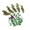
| ||||||||
| Unit cell |
|
-Components
| #1: Protein | Mass: 16237.823 Da / Num. of mol.: 2 Source method: isolated from a genetically manipulated source Source: (gene. exp.) #2: Protein | Mass: 16488.320 Da / Num. of mol.: 2 Source method: isolated from a genetically manipulated source Source: (gene. exp.) #3: Chemical | ChemComp-CMS / |   Mass: 132.118 Da / Num. of mol.: 1 / Source method: obtained synthetically / Formula: C4H8N2O3 Mass: 132.118 Da / Num. of mol.: 1 / Source method: obtained synthetically / Formula: C4H8N2O3#4: Chemical |   Mass: 767.534 Da / Num. of mol.: 2 / Source method: obtained synthetically / Formula: C21H36N7O16P3S Mass: 767.534 Da / Num. of mol.: 2 / Source method: obtained synthetically / Formula: C21H36N7O16P3S#5: Water | ChemComp-HOH / |  Mass: 18.015 Da / Num. of mol.: 276 / Source method: isolated from a natural source / Formula: H2O Mass: 18.015 Da / Num. of mol.: 276 / Source method: isolated from a natural source / Formula: H2O |
|---|
-Experimental details
-Experiment
| Experiment | Method: X-RAY DIFFRACTION / Number of used crystals: 1 |
|---|
- Sample preparation
| Crystal | Density Matthews: 2.42 Å3/Da / Density % sol: 49.12 % |
|---|---|
| Crystal grow | Temperature: 292 K / pH: 6 Details: 0.2 M LiCl 0.1 M MES, PEG 6000, VAPOR DIFFUSION, SITTING DROP, temperature 292K |
-Data collection
| Diffraction | Mean temperature: 100 K |
|---|---|
| Diffraction source | Source: SYNCHROTRON / Site: ESRF  / Beamline: ID14-1 / Wavelength: 0.9334 / Beamline: ID14-1 / Wavelength: 0.9334 |
| Detector | Type: ADSC QUANTUM 210 / Detector: CCD / Date: May 11, 2009 |
| Radiation | Monochromator: GRAPHITE / Protocol: SINGLE WAVELENGTH / Monochromatic (M) / Laue (L): M / Scattering type: x-ray |
| Radiation wavelength | Wavelength: 0.9334 Å / Relative weight: 1 |
| Reflection | Resolution: 2→37.06 Å / Num. obs: 40222 / % possible obs: 98 % / Observed criterion σ(I): 0 |
- Processing
| Software |
| |||||||||||||||||||||||||||||||||||||||||||||||||||||||||||||||||||||||||||||||||||||||||||||||||||||||||||||||||||||||||||||
|---|---|---|---|---|---|---|---|---|---|---|---|---|---|---|---|---|---|---|---|---|---|---|---|---|---|---|---|---|---|---|---|---|---|---|---|---|---|---|---|---|---|---|---|---|---|---|---|---|---|---|---|---|---|---|---|---|---|---|---|---|---|---|---|---|---|---|---|---|---|---|---|---|---|---|---|---|---|---|---|---|---|---|---|---|---|---|---|---|---|---|---|---|---|---|---|---|---|---|---|---|---|---|---|---|---|---|---|---|---|---|---|---|---|---|---|---|---|---|---|---|---|---|---|---|---|---|
| Refinement | Method to determine structure: MOLECULAR REPLACEMENT Starting model: PDB ENTRIES 2D59, 1MJO Resolution: 2→37.06 Å / SU ML: 0.29 / σ(F): 1.98 / Phase error: 24.97 / Stereochemistry target values: ML
| |||||||||||||||||||||||||||||||||||||||||||||||||||||||||||||||||||||||||||||||||||||||||||||||||||||||||||||||||||||||||||||
| Solvent computation | Shrinkage radii: 0.9 Å / VDW probe radii: 1.11 Å / Solvent model: FLAT BULK SOLVENT MODEL / Bsol: 41.45 Å2 / ksol: 0.34 e/Å3 | |||||||||||||||||||||||||||||||||||||||||||||||||||||||||||||||||||||||||||||||||||||||||||||||||||||||||||||||||||||||||||||
| Displacement parameters |
| |||||||||||||||||||||||||||||||||||||||||||||||||||||||||||||||||||||||||||||||||||||||||||||||||||||||||||||||||||||||||||||
| Refinement step | Cycle: LAST / Resolution: 2→37.06 Å
| |||||||||||||||||||||||||||||||||||||||||||||||||||||||||||||||||||||||||||||||||||||||||||||||||||||||||||||||||||||||||||||
| Refine LS restraints |
| |||||||||||||||||||||||||||||||||||||||||||||||||||||||||||||||||||||||||||||||||||||||||||||||||||||||||||||||||||||||||||||
| LS refinement shell |
| |||||||||||||||||||||||||||||||||||||||||||||||||||||||||||||||||||||||||||||||||||||||||||||||||||||||||||||||||||||||||||||
| Refinement TLS params. | Method: refined / Refine-ID: X-RAY DIFFRACTION
| |||||||||||||||||||||||||||||||||||||||||||||||||||||||||||||||||||||||||||||||||||||||||||||||||||||||||||||||||||||||||||||
| Refinement TLS group |
|
