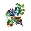
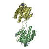
ATP-dependent protein folding chaperone / : / cellular response to heat / protein folding / protein stabilization / perinuclear region of cytoplasm / ATP hydrolysis activity / protein-containing complex / nucleoplasm / ATP binding ...ATP-dependent protein folding chaperone / : / cellular response to heat / protein folding / protein stabilization / perinuclear region of cytoplasm / ATP hydrolysis activity / protein-containing complex / nucleoplasm / ATP binding / metal ion binding / plasma membrane / cytosol / cytoplasm Similarity search - Function
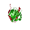
Heat shock protein Hsp90, conserved site / Heat shock hsp90 proteins family signature. / Histidine kinase-like ATPase, C-terminal domain / Heat Shock Protein 90 / HSP90, C-terminal domain / Heat shock protein Hsp90, N-terminal / Heat shock protein Hsp90 family / Hsp90 protein / Histidine kinase-, DNA gyrase B-, and HSP90-like ATPase / Histidine kinase-like ATPases ...Heat shock protein Hsp90, conserved site / Heat shock hsp90 proteins family signature. / Histidine kinase-like ATPase, C-terminal domain / Heat Shock Protein 90 / HSP90, C-terminal domain / Heat shock protein Hsp90, N-terminal / Heat shock protein Hsp90 family / Hsp90 protein / Histidine kinase-, DNA gyrase B-, and HSP90-like ATPase / Histidine kinase-like ATPases / Histidine kinase/HSP90-like ATPase / Histidine kinase/HSP90-like ATPase superfamily / Ribosomal protein S5 domain 2-type fold / 2-Layer Sandwich / Alpha Beta Similarity search - Domain/homology
Resolution: 2.65→24.54 Å / Cor.coef. Fo:Fc: 0.941 / Cor.coef. Fo:Fc free: 0.916 / WRfactor Rfree: 0.2795 / WRfactor Rwork: 0.2239 / Occupancy max: 1 / Occupancy min: 1 / FOM work R set: 0.8043 / SU B: 25.295 / SU ML: 0.242 / SU R Cruickshank DPI: 0.5494 / SU Rfree: 0.3276 / Cross valid method: THROUGHOUT / σ(F): 0 / ESU R Free: 0.328 / Stereochemistry target values: MAXIMUM LIKELIHOOD Details: HYDROGENS HAVE BEEN ADDED IN THE RIDING POSITIONS. U VALUES WITH TLS ADDED.
Rfactor
Num. reflection
% reflection
Selection details
Rfree
0.2777
1822
5 %
RANDOM
Rwork
0.2267
-
-
-
all
0.2292
36598
-
-
obs
0.2292
36452
99.44 %
-
Solvent computation
Ion probe radii: 0.8 Å / Shrinkage radii: 0.8 Å / VDW probe radii: 1.4 Å / Solvent model: MASK
 Movie
Movie Controller

 Yorodumi
 Open data
 Basic information
 Components
 Keywords
 Function and homology information
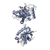
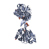
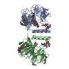
 Leishmania major (eukaryote)
 X-RAY DIFFRACTION /
  Authors
 Citation
 Structure visualization
 Downloads & links
 Other downloads
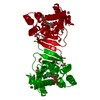


 PDBj
PDBj

 Assembly
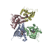


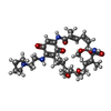
 Mass: 642.783 Da / Num. of mol.: 4 / Source method: obtained synthetically / Formula: C34H50N4O8
 Sample preparation
 / Beamline: 19-ID / Wavelength: 0.97942 Å
 Processing