 Movie
Movie Controller

[English] 日本語
 Yorodumi
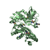
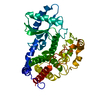
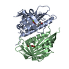
- PDB-3q4n: Crystal structure of hypothetical protein MJ0754 from Methanococc... -
+ Open data

- Basic information
| Entry | Database: PDB / ID: 3q4n | ||||||
|---|---|---|---|---|---|---|---|
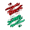
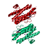
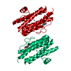
| Title | Crystal structure of hypothetical protein MJ0754 from Methanococcus jannaschii DSM 2661 | ||||||
 Components Components | Uncharacterized protein MJ0754 | ||||||
 Keywords Keywords | UNKNOWN FUNCTION / ferritin-like protein / four-helix bundle / metal binding / dinuclear center | ||||||
| Function / homology | Domain of unknown function DUF2202 / Uncharacterized protein domain (DUF2202) / Ferritin, core subunit, four-helix bundle / Ferritin / Ferritin-like / Ferritin-like superfamily / Up-down Bundle / Mainly Alpha / Uncharacterized protein MJ0754 Function and homology information Function and homology information | ||||||
| Biological species |   Methanocaldococcus jannaschii (archaea) Methanocaldococcus jannaschii (archaea) | ||||||
| Method |  X-RAY DIFFRACTION / SYNCHROTRON / SAD / Resolution: 2.88 Å X-RAY DIFFRACTION / SYNCHROTRON / SAD / Resolution: 2.88 Å | ||||||
 Authors Authors | Hwang, K.Y. / Lee, E.H. | ||||||
 Citation Citation | Journal: Proteins / Year: 2011 Title: Structural insights into the metal binding properties of hypothetical protein MJ0754 from Methanococcus jannaschii. Authors: Lee, E.H. / Kim, H.S. / Kim, H.Y. / Jeon, Y.H. / Hwang, K.Y. | ||||||
| History |
|
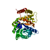
- Structure visualization
| Structure viewer | Molecule: MolmilJmol/JSmol |
|---|
- Downloads & links
-Download
| PDBx/mmCIF format | 3q4n.cif.gz | 79.4 KB | Display | PDBx/mmCIF format |
|---|---|---|---|---|
| PDB format | pdb3q4n.ent.gz | 61.3 KB | Display | PDB format |
| PDBx/mmJSON format | 3q4n.json.gz | Tree view | PDBx/mmJSON format | |
| Others |  Other downloads Other downloads |
-Validation report
| Arichive directory | https://data.pdbj.org/pub/pdb/validation_reports/q4/3q4nftp://data.pdbj.org/pub/pdb/validation_reports/q4/3q4n | HTTPS FTP |
|---|
-Related structure data


-Links
 PDBj
PDBj


- Assembly
| Deposited unit | 
| ||||||||
|---|---|---|---|---|---|---|---|---|---|
| 1 |
| ||||||||
| Unit cell |
|
-Components
| #1: Protein | Mass: 22865.801 Da / Num. of mol.: 2 Source method: isolated from a genetically manipulated source Source: (gene. exp.) Methanocaldococcus jannaschii (archaea)Strain: DSM 2661 / Gene: MJ0754 / Plasmid: pET22b / Production host:  |
|---|
-Experimental details
-Experiment
| Experiment | Method: X-RAY DIFFRACTION / Number of used crystals: 1 |
|---|
- Sample preparation
| Crystal | Density Matthews: 2.49 Å3/Da / Density % sol: 50.63 % |
|---|---|
| Crystal grow | Temperature: 295 K / Method: vapor diffusion, hanging drop / pH: 6 Details: 0.1M Bis-Tris, 22% PEG 3350, pH 6, VAPOR DIFFUSION, HANGING DROP, temperature 295K |
-Data collection
| Diffraction | Mean temperature: 100 K | ||||||||||||||||||||||||||||||||||||||||||||||||||||||||||||||||||
|---|---|---|---|---|---|---|---|---|---|---|---|---|---|---|---|---|---|---|---|---|---|---|---|---|---|---|---|---|---|---|---|---|---|---|---|---|---|---|---|---|---|---|---|---|---|---|---|---|---|---|---|---|---|---|---|---|---|---|---|---|---|---|---|---|---|---|---|
| Diffraction source | Source: SYNCHROTRON / Site: Photon Factory  / Beamline: BL-17A / Wavelength: 0.9795 Å / Beamline: BL-17A / Wavelength: 0.9795 Å | ||||||||||||||||||||||||||||||||||||||||||||||||||||||||||||||||||
| Detector | Type: ADSC QUANTUM 270 / Detector: CCD / Date: Jan 20, 2008 | ||||||||||||||||||||||||||||||||||||||||||||||||||||||||||||||||||
| Radiation | Monochromator: TCM Si(111) / Protocol: SINGLE WAVELENGTH / Monochromatic (M) / Laue (L): M / Scattering type: x-ray | ||||||||||||||||||||||||||||||||||||||||||||||||||||||||||||||||||
| Radiation wavelength | Wavelength: 0.9795 Å / Relative weight: 1 | ||||||||||||||||||||||||||||||||||||||||||||||||||||||||||||||||||
| Reflection | Resolution: 2.88→50 Å / Num. obs: 9543 / % possible obs: 91.8 % / Rmerge(I) obs: 0.147 / Net I/σ(I): 10.3 | ||||||||||||||||||||||||||||||||||||||||||||||||||||||||||||||||||
| Reflection shell |
|
- Processing
| Software |
| |||||||||||||||||||||||||||||||||||||||||||||||||||||||||||||||||
|---|---|---|---|---|---|---|---|---|---|---|---|---|---|---|---|---|---|---|---|---|---|---|---|---|---|---|---|---|---|---|---|---|---|---|---|---|---|---|---|---|---|---|---|---|---|---|---|---|---|---|---|---|---|---|---|---|---|---|---|---|---|---|---|---|---|---|
| Refinement | Method to determine structure: SAD / Resolution: 2.88→31.75 Å / Cor.coef. Fo:Fc: 0.947 / Cor.coef. Fo:Fc free: 0.919 / Occupancy max: 1 / Occupancy min: 1 / SU B: 20.195 / SU ML: 0.378 / Cross valid method: THROUGHOUT / σ(F): 0 / ESU R Free: 0.435 / Stereochemistry target values: MAXIMUM LIKELIHOOD Details: HYDROGENS HAVE BEEN ADDED IN THE RIDING POSITIONS U VALUES: REFINED INDIVIDUALLY
| |||||||||||||||||||||||||||||||||||||||||||||||||||||||||||||||||
| Solvent computation | Ion probe radii: 0.8 Å / Shrinkage radii: 0.8 Å / VDW probe radii: 1.4 Å / Solvent model: MASK | |||||||||||||||||||||||||||||||||||||||||||||||||||||||||||||||||
| Displacement parameters | Biso max: 104.63 Å2 / Biso mean: 75.9443 Å2 / Biso min: 51.22 Å2
| |||||||||||||||||||||||||||||||||||||||||||||||||||||||||||||||||
| Refinement step | Cycle: LAST / Resolution: 2.88→31.75 Å
| |||||||||||||||||||||||||||||||||||||||||||||||||||||||||||||||||
| Refine LS restraints |
| |||||||||||||||||||||||||||||||||||||||||||||||||||||||||||||||||
| LS refinement shell | Resolution: 2.884→2.958 Å / Total num. of bins used: 20
|
