 Movie
Movie Controller

[English] 日本語
 Yorodumi
- PDB-3pvf: Structure of C126S mutant of Plasmodium falciparum triosephosphat... -
+ Open data

- Basic information
| Entry | Database: PDB / ID: 3pvf | ||||||
|---|---|---|---|---|---|---|---|
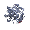
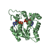
| Title | Structure of C126S mutant of Plasmodium falciparum triosephosphate isomerase complexed with PGA | ||||||
 Components Components | Triosephosphate isomerase | ||||||
 Keywords Keywords | ISOMERASE / Tim barrel | ||||||
| Function / homology |  Function and homology information Function and homology informationtriose-phosphate isomerase / triose-phosphate isomerase activity / glyceraldehyde-3-phosphate biosynthetic process / glycerol catabolic process / gluconeogenesis / glycolytic process / identical protein binding / cytosol Similarity search - Function | ||||||
| Biological species |  | ||||||
| Method |  X-RAY DIFFRACTION / MOLECULAR REPLACEMENT / Resolution: 1.73 Å X-RAY DIFFRACTION / MOLECULAR REPLACEMENT / Resolution: 1.73 Å | ||||||
 Authors Authors | Samanta, M. / Banerjee, M. / Murthy, M.R.N. / Balaram, H. / Balaram, P. | ||||||
 Citation Citation | Journal: Febs J. / Year: 2011 Title: Probing the role of the fully conserved Cys126 in triosephosphate isomerase by site-specific mutagenesis--distal effects on dimer stability. Authors: Samanta, M. / Banerjee, M. / Murthy, M.R. / Balaram, H. / Balaram, P. | ||||||
| History |
|
- Structure visualization
| Structure viewer | Molecule: MolmilJmol/JSmol |
|---|
- Downloads & links
-Download
| PDBx/mmCIF format | 3pvf.cif.gz | 71 KB | Display | PDBx/mmCIF format |
|---|---|---|---|---|
| PDB format | pdb3pvf.ent.gz | 51.4 KB | Display | PDB format |
| PDBx/mmJSON format | 3pvf.json.gz | Tree view | PDBx/mmJSON format | |
| Others |  Other downloads Other downloads |
-Validation report
| Arichive directory | https://data.pdbj.org/pub/pdb/validation_reports/pv/3pvfftp://data.pdbj.org/pub/pdb/validation_reports/pv/3pvf | HTTPS FTP |
|---|
-Related structure data
| Related structure data |  3pwaC  3py2C  1lyxS S: Starting model for refinement C: citing same article ( |
|---|---|
| Similar structure data |


-Links
 PDBj
PDBj
- Assembly
| Deposited unit | 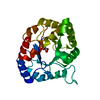
| ||||||||
|---|---|---|---|---|---|---|---|---|---|
| 1 |
| ||||||||
| 2 | 
| ||||||||
| Unit cell |
|
-Components
| #1: Protein | Mass: 27981.674 Da / Num. of mol.: 1 / Mutation: C126S Source method: isolated from a genetically manipulated source Source: (gene. exp.) Gene: TPI / Plasmid: pTrc99A / Production host:  |
|---|---|
| #2: Chemical | ChemComp-PGA /   Mass: 156.031 Da / Num. of mol.: 1 Mass: 156.031 Da / Num. of mol.: 1Source method: isolated from a genetically manipulated source Formula: C2H5O6P |
| #3: Water | ChemComp-HOH /  Mass: 18.015 Da / Num. of mol.: 343 / Source method: isolated from a natural source / Formula: H2O Mass: 18.015 Da / Num. of mol.: 343 / Source method: isolated from a natural source / Formula: H2O |
-Experimental details
-Experiment
| Experiment | Method: X-RAY DIFFRACTION / Number of used crystals: 1 |
|---|
- Sample preparation
| Crystal | Density Matthews: 2.33 Å3/Da / Density % sol: 47.29 % |
|---|---|
| Crystal grow | Temperature: 298 K / Method: vapor diffusion, hanging drop / pH: 7.5 Details: 20% PEG, HEPES buffer(pH 7.5), 10mM Lithium Sulfate, VAPOR DIFFUSION, HANGING DROP, temperature 298K |
-Data collection
| Diffraction | Mean temperature: 100 K |
|---|---|
| Diffraction source | Source: ROTATING ANODE / Type: BRUKER AXS MICROSTAR / Wavelength: 1.541 Å |
| Detector | Type: MAR scanner 345 mm plate / Detector: IMAGE PLATE / Date: Nov 30, 2009 / Details: mirror |
| Radiation | Monochromator: graphite / Protocol: SINGLE WAVELENGTH / Monochromatic (M) / Laue (L): M / Scattering type: x-ray |
| Radiation wavelength | Wavelength: 1.541 Å / Relative weight: 1 |
| Reflection | Resolution: 1.73→38.91 Å / Num. obs: 25341 / % possible obs: 93.94 % / Observed criterion σ(F): 0 / Observed criterion σ(I): 0 / Redundancy: 4.13 % / Rmerge(I) obs: 0.031 / Net I/σ(I): 17.37 |
| Reflection shell | Resolution: 1.732→1.777 Å / Rmerge(I) obs: 0.125 / % possible all: 83.2 |
- Processing
| Software |
| |||||||||||||||||||||||||||||||||||||||||||||||||||||||||||||||||
|---|---|---|---|---|---|---|---|---|---|---|---|---|---|---|---|---|---|---|---|---|---|---|---|---|---|---|---|---|---|---|---|---|---|---|---|---|---|---|---|---|---|---|---|---|---|---|---|---|---|---|---|---|---|---|---|---|---|---|---|---|---|---|---|---|---|---|
| Refinement | Method to determine structure: MOLECULAR REPLACEMENT Starting model: 1LYX Resolution: 1.73→26.25 Å / Cor.coef. Fo:Fc: 0.959 / Cor.coef. Fo:Fc free: 0.936 / SU B: 1.991 / SU ML: 0.065 / Cross valid method: THROUGHOUT / σ(F): 0 / ESU R Free: 0.109 / Stereochemistry target values: MAXIMUM LIKELIHOOD / Details: HYDROGENS HAVE BEEN ADDED IN THE RIDING POSITIONS
| |||||||||||||||||||||||||||||||||||||||||||||||||||||||||||||||||
| Solvent computation | Ion probe radii: 0.8 Å / Shrinkage radii: 0.8 Å / VDW probe radii: 1.4 Å / Solvent model: MASK | |||||||||||||||||||||||||||||||||||||||||||||||||||||||||||||||||
| Displacement parameters | Biso mean: 10.673 Å2
| |||||||||||||||||||||||||||||||||||||||||||||||||||||||||||||||||
| Refinement step | Cycle: LAST / Resolution: 1.73→26.25 Å
| |||||||||||||||||||||||||||||||||||||||||||||||||||||||||||||||||
| Refine LS restraints |
| |||||||||||||||||||||||||||||||||||||||||||||||||||||||||||||||||
| LS refinement shell | Resolution: 1.732→1.777 Å / Total num. of bins used: 20
|
