| Entry | Database: PDB / ID: 3pto
|
|---|
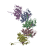
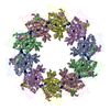
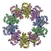
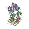
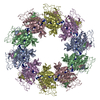
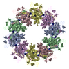
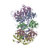
| Title | Crystal Structure of an empty Vesicular Stomatitis Virus Nucleocapsid Protein Complex |
|---|
 Components Components | Nucleoprotein |
|---|
 Keywords Keywords | VIRAL PROTEIN / nucleocapsid |
|---|
| Function / homology |  Function and homology information Function and homology information
RNA replication / helical viral capsid / viral transcription / viral nucleocapsid / host cell cytoplasm / ribonucleoprotein complex / RNA bindingSimilarity search - Function Rhabdovirus nucleoprotein-like / Nucleoprotein / Rhabdovirus nucleoprotein-like fold / Rhabdovirus nucleocapsid protein like domain / Rhabdovirus nucleocapsid / Rhabdovirus nucleocapsid, N-terminal / Rhabdovirus nucleocapsid, C-terminal / Rhabdovirus nucleoprotein-like / Rhabdovirus nucleocapsid protein / Orthogonal Bundle / Mainly AlphaSimilarity search - Domain/homology |
|---|
| Biological species |  Vesicular stomatitis Indiana virus Vesicular stomatitis Indiana virus |
|---|
| Method |  X-RAY DIFFRACTION / SYNCHROTRON / MOLECULAR REPLACEMENT / Resolution: 3.008 Å X-RAY DIFFRACTION / SYNCHROTRON / MOLECULAR REPLACEMENT / Resolution: 3.008 Å |
|---|
 Authors Authors | Luo, M. / Green, T.J. / Rowse, M. |
|---|
 Citation Citation | Journal: J.Virol. / Year: 2011
Title: Access to RNA Encapsidated in the Nucleocapsid of Vesicular Stomatitis Virus.
Authors: Green, T.J. / Rowse, M. / Tsao, J. / Kang, J. / Ge, P. / Zhou, Z.H. / Luo, M. |
|---|
| History | | Deposition | Dec 3, 2010 | Deposition site: RCSB / Processing site: RCSB |
|---|
| Revision 1.0 | Jan 12, 2011 | Provider: repository / Type: Initial release |
|---|
| Revision 1.1 | Jul 13, 2011 | Group: Version format compliance |
|---|
| Revision 1.2 | Feb 21, 2024 | Group: Data collection / Database references / Derived calculations
Category: chem_comp_atom / chem_comp_bond ...chem_comp_atom / chem_comp_bond / database_2 / pdbx_struct_conn_angle / struct_conn / struct_site
Item: _database_2.pdbx_DOI / _database_2.pdbx_database_accession ..._database_2.pdbx_DOI / _database_2.pdbx_database_accession / _pdbx_struct_conn_angle.ptnr1_auth_asym_id / _pdbx_struct_conn_angle.ptnr1_auth_comp_id / _pdbx_struct_conn_angle.ptnr1_auth_seq_id / _pdbx_struct_conn_angle.ptnr1_label_asym_id / _pdbx_struct_conn_angle.ptnr1_label_atom_id / _pdbx_struct_conn_angle.ptnr1_label_comp_id / _pdbx_struct_conn_angle.ptnr1_label_seq_id / _pdbx_struct_conn_angle.ptnr2_auth_asym_id / _pdbx_struct_conn_angle.ptnr2_auth_seq_id / _pdbx_struct_conn_angle.ptnr2_label_asym_id / _pdbx_struct_conn_angle.ptnr3_auth_asym_id / _pdbx_struct_conn_angle.ptnr3_auth_comp_id / _pdbx_struct_conn_angle.ptnr3_auth_seq_id / _pdbx_struct_conn_angle.ptnr3_label_asym_id / _pdbx_struct_conn_angle.ptnr3_label_atom_id / _pdbx_struct_conn_angle.ptnr3_label_comp_id / _pdbx_struct_conn_angle.ptnr3_label_seq_id / _pdbx_struct_conn_angle.value / _struct_conn.pdbx_dist_value / _struct_conn.ptnr1_auth_asym_id / _struct_conn.ptnr1_auth_comp_id / _struct_conn.ptnr1_auth_seq_id / _struct_conn.ptnr1_label_asym_id / _struct_conn.ptnr1_label_atom_id / _struct_conn.ptnr1_label_comp_id / _struct_conn.ptnr1_label_seq_id / _struct_conn.ptnr2_auth_asym_id / _struct_conn.ptnr2_auth_comp_id / _struct_conn.ptnr2_auth_seq_id / _struct_conn.ptnr2_label_asym_id / _struct_conn.ptnr2_label_atom_id / _struct_conn.ptnr2_label_comp_id / _struct_conn.ptnr2_label_seq_id / _struct_site.pdbx_auth_asym_id / _struct_site.pdbx_auth_comp_id / _struct_site.pdbx_auth_seq_id |
|---|
|
|---|
 Movie
Movie Controller

 Yorodumi
 Open data
 Basic information
 Structure visualization
 Downloads & links
 Other downloads


 PDBj
PDBj

 Assembly