Mass: 1379.345 Da / Num. of mol.: 2 / Source method: obtained synthetically Details: This is a designed peptide (two residues switched position) based on human Met protein sequence.
#2: Protein
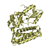
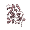
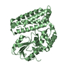
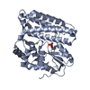
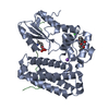
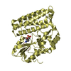
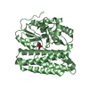
E3ubiquitin-proteinligaseCBL / Casitas B-lineage lymphoma proto-oncogene / Proto-oncogene c-Cbl / RING finger protein 55 / Signal ...Casitas B-lineage lymphoma proto-oncogene / Proto-oncogene c-Cbl / RING finger protein 55 / Signal transduction protein CBL
Mass: 38192.090 Da / Num. of mol.: 2 / Fragment: TKB domain Source method: isolated from a genetically manipulated source Source: (gene. exp.) Homo sapiens (human) / Gene: c-Cbl / Plasmid: pGEX-4T-1 / Production host: Escherichia coli (E. coli) / Strain (production host): BL21(DE3) References: UniProt: P22681, Ligases; Forming carbon-nitrogen bonds; Acid-amino-acid ligases (peptide synthases)
Resolution: 1.92→47.08 Å / Cor.coef. Fo:Fc: 0.961 / Cor.coef. Fo:Fc free: 0.949 / WRfactor Rfree: 0.1889 / WRfactor Rwork: 0.1681 / Occupancy max: 1 / Occupancy min: 1 / FOM work R set: 0.9004 / SU B: 5.31 / SU ML: 0.071 / SU R Cruickshank DPI: 0.1097 / SU Rfree: 0.1242 / Cross valid method: THROUGHOUT / σ(F): 0 / ESU R Free: 0.124 / Stereochemistry target values: MAXIMUM LIKELIHOOD Details: HYDROGENS HAVE BEEN ADDED IN THE RIDING POSITIONS U VALUES: RESIDUAL ONLY
Rfactor
Num. reflection
% reflection
Selection details
Rfree
0.1802
2462
5.1 %
RANDOM
Rwork
0.1591
-
-
-
all
0.1602
-
-
-
obs
0.1602
46063
97.4 %
-
Solvent computation
Ion probe radii: 0.8 Å / Shrinkage radii: 0.8 Å / VDW probe radii: 1.4 Å / Solvent model: MASK
In the structure databanks used in Yorodumi, some data are registered as the other names, "COVID-19 virus" and "2019-nCoV". Here are the details of the virus and the list of structure data.
Jan 31, 2019. EMDB accession codes are about to change! (news from PDBe EMDB page)
EMDB accession codes are about to change! (news from PDBe EMDB page)
The allocation of 4 digits for EMDB accession codes will soon come to an end. Whilst these codes will remain in use, new EMDB accession codes will include an additional digit and will expand incrementally as the available range of codes is exhausted. The current 4-digit format prefixed with “EMD-” (i.e. EMD-XXXX) will advance to a 5-digit format (i.e. EMD-XXXXX), and so on. It is currently estimated that the 4-digit codes will be depleted around Spring 2019, at which point the 5-digit format will come into force.
The EM Navigator/Yorodumi systems omit the EMD- prefix.
Related info.:Q: What is EMD? / ID/Accession-code notation in Yorodumi/EM Navigator
Yorodumi is a browser for structure data from EMDB, PDB, SASBDB, etc.
This page is also the successor to EM Navigator detail page, and also detail information page/front-end page for Omokage search.
The word "yorodu" (or yorozu) is an old Japanese word meaning "ten thousand". "mi" (miru) is to see.
Related info.:EMDB / PDB / SASBDB / Comparison of 3 databanks / Yorodumi Search / Aug 31, 2016. New EM Navigator & Yorodumi / Yorodumi Papers / Jmol/JSmol / Function and homology information / Changes in new EM Navigator and Yorodumi
 Movie
Movie Controller

 Yorodumi
 Open data
 Basic information
 Components
 Keywords
 Function and homology information
 Homo sapiens (human)
 X-RAY DIFFRACTION /
  Authors
 Citation
 Structure visualization
 Downloads & links
 Other downloads
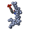


 PDBj
PDBj


 Assembly


 Mass: 40.078 Da / Num. of mol.: 2 / Source method: obtained synthetically / Formula: Ca
 Mass: 35.453 Da / Num. of mol.: 4 / Source method: obtained synthetically / Formula: Cl
 Mass: 18.015 Da / Num. of mol.: 584 / Source method: isolated from a natural source / Formula: H2O
 Sample preparation
 Processing