 Movie
Movie Controller

[English] 日本語
 Yorodumi
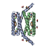
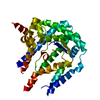
- PDB-3pl9: Crystal structure of spinach minor light-harvesting complex CP29 ... -
+ Open data

- Basic information
| Entry | Database: PDB / ID: 3pl9 | ||||||||||||
|---|---|---|---|---|---|---|---|---|---|---|---|---|---|
| Title | Crystal structure of spinach minor light-harvesting complex CP29 at 2.80 angstrom resolution | ||||||||||||
 Components Components | Chlorophyll A-B binding protein | ||||||||||||
 Keywords Keywords | PHOTOSYNTHESIS / CP29 / Chlorophyll A-B binding protein / light-harvesting complex / membrane protein / plant / chloroplast / thylakoid / photosystem II | ||||||||||||
| Function / homology |  Function and homology information Function and homology informationphotosynthesis, light harvesting / photosystem I / photosystem II / chlorophyll binding / chloroplast thylakoid membrane / phosphoprotein binding / mRNA binding / metal ion binding Similarity search - Function | ||||||||||||
| Biological species |  Spinacia oleracea (spinach) Spinacia oleracea (spinach) | ||||||||||||
| Method |  X-RAY DIFFRACTION / SYNCHROTRON / SIRAS / Resolution: 2.8 Å X-RAY DIFFRACTION / SYNCHROTRON / SIRAS / Resolution: 2.8 Å | ||||||||||||
 Authors Authors | Pan, X.W. / Li, M. / Wan, T. / Wang, L.F. / Jia, C.J. / Hou, Z.Q. / Zhao, X.L. / Zhang, J.P. / Chang, W.R. | ||||||||||||
 Citation Citation | Journal: Nat.Struct.Mol.Biol. / Year: 2011 Title: Structural insights into energy regulation of light-harvesting complex CP29 from spinach. Authors: Pan, X. / Li, M. / Wan, T. / Wang, L. / Jia, C. / Hou, Z. / Zhao, X. / Zhang, J. / Chang, W. | ||||||||||||
| History |
|
- Structure visualization
| Structure viewer | Molecule: MolmilJmol/JSmol |
|---|
- Downloads & links
-Download
| PDBx/mmCIF format | 3pl9.cif.gz | 79.9 KB | Display | PDBx/mmCIF format |
|---|---|---|---|---|
| PDB format | pdb3pl9.ent.gz | 60.5 KB | Display | PDB format |
| PDBx/mmJSON format | 3pl9.json.gz | Tree view | PDBx/mmJSON format | |
| Others |  Other downloads Other downloads |
-Validation report
| Arichive directory | https://data.pdbj.org/pub/pdb/validation_reports/pl/3pl9ftp://data.pdbj.org/pub/pdb/validation_reports/pl/3pl9 | HTTPS FTP |
|---|
-Related structure data
| Similar structure data |
|---|


-Links
 PDBj
PDBj


- Assembly
| Deposited unit | 
| ||||||||
|---|---|---|---|---|---|---|---|---|---|
| 1 |
| ||||||||
| Unit cell |
|
-Components
-Protein / Sugars , 2 types, 3 molecules A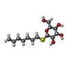
| #1: Protein | Mass: 26735.303 Da / Num. of mol.: 1 / Source method: isolated from a natural source / Source: (natural) Spinacia oleracea (spinach) / References: UniProt: F2Z293*PLUS |
|---|---|
| #8: Sugar |  Type: D-saccharide / Mass: 294.408 Da / Num. of mol.: 2 / Source method: obtained synthetically / Formula: C13H26O5S / Comment: detergent*YM Type: D-saccharide / Mass: 294.408 Da / Num. of mol.: 2 / Source method: obtained synthetically / Formula: C13H26O5S / Comment: detergent*YM |
-Non-polymers , 7 types, 96 molecules 

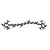
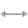


| #2: Chemical | ChemComp-CLA /  Mass: 893.489 Da / Num. of mol.: 9 / Source method: obtained synthetically / Formula: C55H72MgN4O5 Mass: 893.489 Da / Num. of mol.: 9 / Source method: obtained synthetically / Formula: C55H72MgN4O5#3: Chemical | ChemComp-CHL /  Mass: 907.472 Da / Num. of mol.: 4 / Source method: obtained synthetically / Formula: C55H70MgN4O6 Mass: 907.472 Da / Num. of mol.: 4 / Source method: obtained synthetically / Formula: C55H70MgN4O6#4: Chemical | ChemComp-LUT / ( |  Mass: 568.871 Da / Num. of mol.: 1 / Source method: obtained synthetically / Formula: C40H56O2 Mass: 568.871 Da / Num. of mol.: 1 / Source method: obtained synthetically / Formula: C40H56O2#5: Chemical | ChemComp-XAT / ( |  Mass: 600.870 Da / Num. of mol.: 1 / Source method: obtained synthetically / Formula: C40H56O4 Mass: 600.870 Da / Num. of mol.: 1 / Source method: obtained synthetically / Formula: C40H56O4#6: Chemical | ChemComp-NEX / ( |  Mass: 600.870 Da / Num. of mol.: 1 / Source method: obtained synthetically / Formula: C40H56O4 Mass: 600.870 Da / Num. of mol.: 1 / Source method: obtained synthetically / Formula: C40H56O4#7: Chemical | ChemComp-G3P / |  Mass: 172.074 Da / Num. of mol.: 1 / Source method: obtained synthetically / Formula: C3H9O6P Mass: 172.074 Da / Num. of mol.: 1 / Source method: obtained synthetically / Formula: C3H9O6P#9: Water | ChemComp-HOH / | Mass: 18.015 Da / Num. of mol.: 79 / Source method: isolated from a natural source / Formula: H2O |
|---|
-Details
| Sequence details | A SEQUENCE DATABASE REFERENCE FOR THIS PROTEIN DOES NOT CURRENTLY EXIST. |
|---|
-Experimental details
-Experiment
| Experiment | Method: X-RAY DIFFRACTION / Number of used crystals: 1 |
|---|
- Sample preparation
| Crystal | Density Matthews: 5.44 Å3/Da / Density % sol: 77.39 % |
|---|---|
| Crystal grow | Temperature: 291 K / Method: vapor diffusion, sitting drop / pH: 6.5 Details: 100mM MES, pH 6.5, 2.80% (w/v) HTG, 19% (w/v) PEG 1000, 0.1M sodium chloride, 0.19M ammonium sulfate , VAPOR DIFFUSION, SITTING DROP, temperature 291K |
-Data collection
| Diffraction | Mean temperature: 100 K |
|---|---|
| Diffraction source | Source: SYNCHROTRON / Site: SSRF  / Beamline: BL17U / Wavelength: 0.99985 Å / Beamline: BL17U / Wavelength: 0.99985 Å |
| Detector | Type: RAYONIX MX225HE / Detector: CCD / Date: Jan 15, 2010 |
| Radiation | Monochromator: SAGITALLY FOCUSED Si(111) / Protocol: SINGLE WAVELENGTH / Monochromatic (M) / Laue (L): M / Scattering type: x-ray |
| Radiation wavelength | Wavelength: 0.99985 Å / Relative weight: 1 |
| Reflection | Resolution: 2.8→50 Å / Num. all: 15959 / Num. obs: 14714 / % possible obs: 92.2 % / Observed criterion σ(F): 0 / Observed criterion σ(I): 0 / Redundancy: 11.9 % / Biso Wilson estimate: 58 Å2 / Rmerge(I) obs: 0.114 / Net I/σ(I): 18.5 |
| Reflection shell | Resolution: 2.8→2.9 Å / Redundancy: 11.2 % / Rmerge(I) obs: 0.586 / Mean I/σ(I) obs: 5.6 / Num. unique all: 627 / % possible all: 40.9 |
- Processing
| Software |
| ||||||||||||||||||||||||||||||||||||||||||||||||||||||||||||||||||||||||||||||||
|---|---|---|---|---|---|---|---|---|---|---|---|---|---|---|---|---|---|---|---|---|---|---|---|---|---|---|---|---|---|---|---|---|---|---|---|---|---|---|---|---|---|---|---|---|---|---|---|---|---|---|---|---|---|---|---|---|---|---|---|---|---|---|---|---|---|---|---|---|---|---|---|---|---|---|---|---|---|---|---|---|---|
| Refinement | Method to determine structure: SIRAS / Resolution: 2.8→42.56 Å / Rfactor Rfree error: 0.011 / Data cutoff high absF: 3581533.95 / Data cutoff low absF: 0 / Isotropic thermal model: RESTRAINED / Cross valid method: THROUGHOUT / σ(F): 0 / σ(I): 0 / Stereochemistry target values: Engh & Huber / Details: BULK SOLVENT MODEL USED
| ||||||||||||||||||||||||||||||||||||||||||||||||||||||||||||||||||||||||||||||||
| Solvent computation | Solvent model: FLAT MODEL / Bsol: 40.4318 Å2 / ksol: 0.25 e/Å3 | ||||||||||||||||||||||||||||||||||||||||||||||||||||||||||||||||||||||||||||||||
| Displacement parameters | Biso mean: 57.2 Å2
| ||||||||||||||||||||||||||||||||||||||||||||||||||||||||||||||||||||||||||||||||
| Refine analyze |
| ||||||||||||||||||||||||||||||||||||||||||||||||||||||||||||||||||||||||||||||||
| Refinement step | Cycle: LAST / Resolution: 2.8→42.56 Å
| ||||||||||||||||||||||||||||||||||||||||||||||||||||||||||||||||||||||||||||||||
| Refine LS restraints |
| ||||||||||||||||||||||||||||||||||||||||||||||||||||||||||||||||||||||||||||||||
| LS refinement shell | Resolution: 2.8→2.98 Å / Rfactor Rfree error: 0.048 / Total num. of bins used: 6
| ||||||||||||||||||||||||||||||||||||||||||||||||||||||||||||||||||||||||||||||||
| Xplor file |
|
