 Movie
Movie Controller

[English] 日本語
 Yorodumi
- PDB-3pl0: Crystal structure of a bsmA homolog (Mpe_A2762) from Methylobium ... -
+ Open data

- Basic information
| Entry | Database: PDB / ID: 3pl0 | ||||||
|---|---|---|---|---|---|---|---|
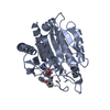
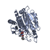
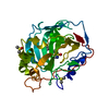
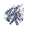
| Title | Crystal structure of a bsmA homolog (Mpe_A2762) from Methylobium petroleophilum PM1 at 1.91 A resolution | ||||||
 Components Components | Uncharacterized protein | ||||||
 Keywords Keywords | BIOSYNTHETIC PROTEIN / QUORUM SENSING / BIOFILM FORMATION / DOUBLE-STRANDED BETA-HELIX FOLD / STRUCTURAL GENOMICS / JOINT CENTER FOR STRUCTURAL GENOMICS / JCSG / PROTEIN STRUCTURE INITIATIVE / PSI-BIOLOGY | ||||||
| Function / homology | 2OG-Fe dioxygenase / 2OG-Fe dioxygenase / q2cbj1_9rhob like domain / dioxygenase activity / Jelly Rolls / Sandwich / Mainly Beta / 2OG-Fe dioxygenase family protein Function and homology information Function and homology information | ||||||
| Biological species |  Methylibium petroleiphilum (bacteria) Methylibium petroleiphilum (bacteria) | ||||||
| Method |  X-RAY DIFFRACTION / SYNCHROTRON / MAD / Resolution: 1.91 Å X-RAY DIFFRACTION / SYNCHROTRON / MAD / Resolution: 1.91 Å | ||||||
 Authors Authors | Joint Center for Structural Genomics (JCSG) | ||||||
 Citation Citation | Journal: Proteins / Year: 2014 Title: Crystal structure of a member of a novel family of dioxygenases (PF10014) reveals a conserved cupin fold and active site. Authors: Xu, Q. / Grant, J. / Chiu, H.J. / Farr, C.L. / Jaroszewski, L. / Knuth, M.W. / Miller, M.D. / Lesley, S.A. / Godzik, A. / Elsliger, M.A. / Deacon, A.M. / Wilson, I.A. | ||||||
| History |
|
- Structure visualization
| Structure viewer | Molecule: MolmilJmol/JSmol |
|---|
- Downloads & links
-Download
| PDBx/mmCIF format | 3pl0.cif.gz | 222.9 KB | Display | PDBx/mmCIF format |
|---|---|---|---|---|
| PDB format | pdb3pl0.ent.gz | 179.8 KB | Display | PDB format |
| PDBx/mmJSON format | 3pl0.json.gz | Tree view | PDBx/mmJSON format | |
| Others |  Other downloads Other downloads |
-Validation report
| Arichive directory | https://data.pdbj.org/pub/pdb/validation_reports/pl/3pl0ftp://data.pdbj.org/pub/pdb/validation_reports/pl/3pl0 | HTTPS FTP |
|---|
-Related structure data
| Similar structure data | |
|---|---|
| Other databases |


-Links
 PDBj
PDBj- Assembly
| Deposited unit | 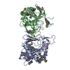
| ||||||||||||||||||
|---|---|---|---|---|---|---|---|---|---|---|---|---|---|---|---|---|---|---|---|
| 1 | 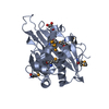
| ||||||||||||||||||
| 2 | 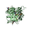
| ||||||||||||||||||
| Unit cell |
| ||||||||||||||||||
| Noncrystallographic symmetry (NCS) | NCS domain:
NCS domain segments: Component-ID: 1 / Ens-ID: 1 / Beg auth comp-ID: GLY / Beg label comp-ID: GLY / End auth comp-ID: ALA / End label comp-ID: ALA / Refine code: 4 / Auth seq-ID: 0 - 253 / Label seq-ID: 1 - 254
|
-Components
| #1: Protein | Mass: 28642.471 Da / Num. of mol.: 2 Source method: isolated from a genetically manipulated source Source: (gene. exp.) Methylibium petroleiphilum (bacteria) / Strain: PM1 / Gene: Mpe_A2762 / Plasmid: SpeedET / Production host: #2: Chemical | ChemComp-GOL / |   Mass: 92.094 Da / Num. of mol.: 1 / Source method: obtained synthetically / Formula: C3H8O3 Mass: 92.094 Da / Num. of mol.: 1 / Source method: obtained synthetically / Formula: C3H8O3#3: Chemical | ChemComp-CL / |   Mass: 35.453 Da / Num. of mol.: 1 / Source method: obtained synthetically / Formula: Cl Mass: 35.453 Da / Num. of mol.: 1 / Source method: obtained synthetically / Formula: Cl#4: Water | ChemComp-HOH / |  Mass: 18.015 Da / Num. of mol.: 413 / Source method: isolated from a natural source / Formula: H2O Mass: 18.015 Da / Num. of mol.: 413 / Source method: isolated from a natural source / Formula: H2OHas protein modification | Y | Sequence details | THE CONSTRUCT WAS EXPRESSED WITH A PURIFICATION TAG MGSDKIHHHHHHENLYFQG. THE TAG WAS REMOVED WITH ...THE CONSTRUCT WAS EXPRESSED WITH A PURIFICATI | |
|---|
-Experimental details
-Experiment
| Experiment | Method: X-RAY DIFFRACTION / Number of used crystals: 1 |
|---|
- Sample preparation
| Crystal | Density Matthews: 2.49 Å3/Da / Density % sol: 50.5 % |
|---|---|
| Crystal grow | Temperature: 277 K / Method: vapor diffusion, sitting drop / pH: 6.5 Details: 0.90M lithium chloride, 6.00% polyethylene glycol 6000, 0.1M HEPES pH 6.5, NANODROP, VAPOR DIFFUSION, SITTING DROP, temperature 277K |
-Data collection
| Diffraction | Mean temperature: 100 K | |||||||||||||||||||||||||||||||||||||||||||||||||||||||||||||||||||||||||||||
|---|---|---|---|---|---|---|---|---|---|---|---|---|---|---|---|---|---|---|---|---|---|---|---|---|---|---|---|---|---|---|---|---|---|---|---|---|---|---|---|---|---|---|---|---|---|---|---|---|---|---|---|---|---|---|---|---|---|---|---|---|---|---|---|---|---|---|---|---|---|---|---|---|---|---|---|---|---|---|
| Diffraction source | Source: SYNCHROTRON / Site: ALS  / Beamline: 8.2.2 / Wavelength: 0.9537,0.9796,0.9793 / Beamline: 8.2.2 / Wavelength: 0.9537,0.9796,0.9793 | |||||||||||||||||||||||||||||||||||||||||||||||||||||||||||||||||||||||||||||
| Detector | Type: ADSC QUANTUM 315 / Detector: CCD / Date: Sep 15, 2010 | |||||||||||||||||||||||||||||||||||||||||||||||||||||||||||||||||||||||||||||
| Radiation | Protocol: MAD / Monochromatic (M) / Laue (L): M / Scattering type: x-ray | |||||||||||||||||||||||||||||||||||||||||||||||||||||||||||||||||||||||||||||
| Radiation wavelength |
| |||||||||||||||||||||||||||||||||||||||||||||||||||||||||||||||||||||||||||||
| Reflection | Resolution: 1.91→28.923 Å / Num. obs: 43643 / % possible obs: 96.6 % / Observed criterion σ(I): -3 / Biso Wilson estimate: 32.356 Å2 / Rmerge(I) obs: 0.042 / Net I/σ(I): 10.4 | |||||||||||||||||||||||||||||||||||||||||||||||||||||||||||||||||||||||||||||
| Reflection shell |
|
-Phasing
| Phasing | Method: MAD |
|---|
- Processing
| Software |
| |||||||||||||||||||||||||||||||||||||||||||||||||||||||||||||||||||||||||||||||||||||
|---|---|---|---|---|---|---|---|---|---|---|---|---|---|---|---|---|---|---|---|---|---|---|---|---|---|---|---|---|---|---|---|---|---|---|---|---|---|---|---|---|---|---|---|---|---|---|---|---|---|---|---|---|---|---|---|---|---|---|---|---|---|---|---|---|---|---|---|---|---|---|---|---|---|---|---|---|---|---|---|---|---|---|---|---|---|---|
| Refinement | Method to determine structure: MAD / Resolution: 1.91→28.923 Å / Cor.coef. Fo:Fc: 0.97 / Cor.coef. Fo:Fc free: 0.956 / Occupancy max: 1 / Occupancy min: 0.3 / SU B: 6.692 / SU ML: 0.098 / Cross valid method: THROUGHOUT / σ(F): 0 / ESU R: 0.145 / ESU R Free: 0.135 Stereochemistry target values: MAXIMUM LIKELIHOOD WITH PHASES Details: 1. HYDROGENS HAVE BEEN ADDED IN THE RIDING POSITIONS. 2. A MET-INHIBITION PROTOCOL WAS USED FOR SELENOMETHIONINE INCORPORATION DURING PROTEIN EXPRESSION. THE OCCUPANCY OF THE SE ATOMS IN THE ...Details: 1. HYDROGENS HAVE BEEN ADDED IN THE RIDING POSITIONS. 2. A MET-INHIBITION PROTOCOL WAS USED FOR SELENOMETHIONINE INCORPORATION DURING PROTEIN EXPRESSION. THE OCCUPANCY OF THE SE ATOMS IN THE MSE RESIDUES WAS REDUCED TO 0.75 FOR THE REDUCED SCATTERING POWER DUE TO PARTIAL S-MET INCORPORATION. 3. GLYCEROL (GOL) AND CHLORIDE (CL) MODELED ARE PRESENT IN CRYO/CRYSTALLIZATION CONDITIONS. 4. ATOM RECORD CONTAINS SUM OF TLS AND RESIDUAL B FACTORS. ANISOU RECORD CONTAINS SUM OF TLS AND RESIDUAL U FACTORS. 5. WATERS WERE EXCLUDED FROM AUTOMATIC TLS ASSIGNMENT.
| |||||||||||||||||||||||||||||||||||||||||||||||||||||||||||||||||||||||||||||||||||||
| Solvent computation | Ion probe radii: 0.8 Å / Shrinkage radii: 0.8 Å / VDW probe radii: 1.2 Å / Solvent model: MASK | |||||||||||||||||||||||||||||||||||||||||||||||||||||||||||||||||||||||||||||||||||||
| Displacement parameters | Biso max: 99.69 Å2 / Biso mean: 35.7943 Å2 / Biso min: 15.87 Å2
| |||||||||||||||||||||||||||||||||||||||||||||||||||||||||||||||||||||||||||||||||||||
| Refinement step | Cycle: LAST / Resolution: 1.91→28.923 Å
| |||||||||||||||||||||||||||||||||||||||||||||||||||||||||||||||||||||||||||||||||||||
| Refine LS restraints |
| |||||||||||||||||||||||||||||||||||||||||||||||||||||||||||||||||||||||||||||||||||||
| Refine LS restraints NCS | Dom-ID: 1 / Auth asym-ID: A / Ens-ID: 1 / Number: 3217 / Refine-ID: X-RAY DIFFRACTION
| |||||||||||||||||||||||||||||||||||||||||||||||||||||||||||||||||||||||||||||||||||||
| LS refinement shell | Resolution: 1.905→1.955 Å / Total num. of bins used: 20
| |||||||||||||||||||||||||||||||||||||||||||||||||||||||||||||||||||||||||||||||||||||
| Refinement TLS params. | Method: refined / Refine-ID: X-RAY DIFFRACTION
| |||||||||||||||||||||||||||||||||||||||||||||||||||||||||||||||||||||||||||||||||||||
| Refinement TLS group |
|
